



Temporal dynamics of serum bilirubin in acute appendicitis: implications for an early hepatobiliary response





Abstract
Background: Hyperbilirubinemia has traditionally been regarded as a marker of complicated acute appendicitis. However, the correlation between serum bilirubin and symptom duration remains poorly described. Our aim was to evaluate the correlation between symptom duration and bilirubin concentrations in patients with histopathologically confirmed acute appendicitis.
Methods: This single-center study included 54 patients with acute appendicitis. Bilirubin fractions, inflammatory markers and other laboratory parameters were analyzed. Their relationship with symptom duration were assessed using Spearman correlation and subgroup comparisons.
Results: Symptom duration inversely correlated with total, direct and indirect bilirubin. Although patients presenting within 24 hours demonstrated numerically higher bilirubin concentrations, subgroup differences did not reach statistical significance.
Conclusion: Serum bilirubin demonstrated a significant inverse association with symptom duration in acute appendicitis, supporting further investigation of bilirubin within a temporal framework.
Citation
Gelevski R, Jota G, Krsteska B, Andreevski V, Kostovska I. Temporal dynamics of serum bilirubin in acute appendicitis: implications for an early hepatobiliary response. Eur J Transl Clin Med.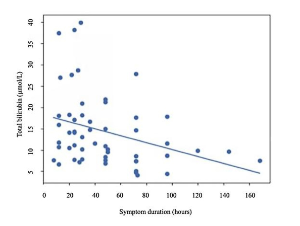








Introduction
Acute appendicitis remains one of the most common surgical emergencies worldwide and a leading indication for urgent abdominal surgery [1-2]. Despite advances in diagnostic imaging and laboratory testing, early identification of biologically active disease and differentiation between uncomplicated and complicated appendicitis continue to pose clinical challenges. Consequently, considerable attention has been directed toward the identification of laboratory biomarkers capable of improving risk stratification and supporting clinical decision-making [3-5].
Multiple studies and meta-analyses have demonstrated an association between hyperbilirubinemia and complicated appendicitis, particularly its gangrenous and perforated forms [6-9]. Within this framework, hyperbilirubinemia has traditionally been interpreted as a marker of advanced disease severity and increased inflammatory burden.
However, most previous investigations have evaluated bilirubin concentration as a static marker measured at a single time point, with relatively little attention paid to its changes during the course of appendicitis. This is an important limitation because the interpretation of any biomarker depends not only on its absolute value but also on the timing of measurement relative to the course of disease. While inflammatory markers such as C-reactive protein (CRP), interleukin-6 (IL-6) and leukocyte-based indices have well-known temporal changes during acute inflammation, the relationship between symptom duration and serum bilirubin concentrations remains insufficiently characterized [10-11].
Our previous clinical observations suggested that elevated bilirubin concentration was frequently encountered in patients presenting relatively early in the course of appendicitis, including some with only modest elevations of conventional inflammatory markers. That raises the possibility that bilirubin may have a different temporal pattern than the traditional inflammatory biomarkers and that it may reflect other biological processes [12].
Several biological mechanisms support this possibility. The liver occupies a central position within the gut–liver axis and is continuously exposed to microbial products delivered through the portal circulation [13-14]. Experimental studies have demonstrated that bacterial endotoxins (particularly lipopolysaccharide) can rapidly alter hepatocellular transporter function and bile excretion before the development of overt systemic inflammatory manifestations [14-15]. These observations provide a biologically plausible basis for early alterations in bilirubin metabolism during intra-abdominal infection. The aim of the present study was to evaluate the correlation between symptom duration and serum bilirubin concentrations in patients with histopathologically confirmed acute appendicitis and to characterize this relationship in the context of inflammatory markers and pathological severity.
Material and methods
This single-center observational study was conducted at the Department of Surgery of the General Hospital in Kumanovo (North Macedonia). Consecutive adult patients undergoing appendectomy for suspected acute appendicitis between July 2025 and December 2025 were screened for inclusion. Only patients with histopathologically confirmed acute appendicitis were included in the final analysis.
Data collection
Clinical and laboratory data were retrospectively collected from electronic medical records. We recorded demographic characteristics (age and sex), symptom duration (defined as the interval between the onset of abdominal pain and surgical intervention, based on patient-reported history) and laboratory parameters (see below).
All laboratory analyses were performed in the central clinical laboratory of the same hospital. Blood samples were obtained at admission, before surgical intervention, and prior to administration of perioperative antibiotics. Complete blood count, including white blood cell count, hemoglobin, hematocrit, and differential leukocyte counts used for calculation of the neutrophil-to-lymphocyte ratio were analyzed using the Beckman Coulter DxH 560 Hematology Analyzer (Beckman Coulter Inc., Brea, CA, USA). Biochemical analyses, including total, direct, and indirect bilirubin, alanine aminotransferase (ALT), aspartate aminotransferase (AST), alkaline phosphatase (ALP), gamma-glutamyl transferase (GGT), creatinine, and urea, were performed using the Atellica® Solution Clinical Chemistry System (Siemens Healthineers AG, Erlangen, Germany). Serum interleukin-6 (IL-6) concentrations were measured using the IMMULITE® 2000 XPi Immunoassay System with the IMMULITE® IL-6 assay (Siemens Healthineers AG, Erlangen, Germany), while serum procalcitonin (PCT) concentrations were determined using the VIDAS® BRAHMS PCT assay on the VIDAS® immunoanalyzer (bioMérieux SA, Marcy-l’Étoile, France). All analyses were performed in the same laboratory, accredited by the National Accreditation Board, following the manufacturers’ instructions for calibration and internal quality-control procedures.
To minimize factors affecting bilirubin metabolism, patients with documented chronic liver disease, viral hepatitis, known Gilbert syndrome, alcohol-related liver disease, biliary tract disease, hemolytic disorders, chronic kidney disease or other conditions known to influence bilirubin metabolism were excluded. Patients with abnormal peripheral blood smear findings suggestive of hemolysis were also excluded. Serological testing for hepatitis (HBsAg, anti-HCV, anti-HAV IgM) was negative in all included patients.
Abdominal ultrasonography was performed in all patients, while computed tomography was obtained when clinically indicated. None of the patients had findings suggestive of clinically significant biliary obstruction or cholestatic disease that could account for the observed bilirubin concentrations. To further exclude previously unrecognized hereditary hyperbilirubinemia, follow-up bilirubin measurements were obtained on postoperative day 30 day.
Definition of pathological severity
Patients were stratified according to histopathological findings into:
- complicated appendicitis (CApp): gangrenous and/or perforated appendicitis,
- uncomplicated appendicitis (non-CApp): phlegmonous or catarrhal appendicitis without evidence of gangrene or perforation.
Study outcomes
Our primary objective was to evaluate the relationship between symptom duration and serum bilirubin concentrations in patients with acute appendicitis. Secondary objectives included:
- assessment of this relationship within subgroups (CApp vs. non-KCApp),
- comparison of laboratory parameters between early and late presentation (≤ 24 h vs. > 24 h),
- evaluation of temporal relationships between symptom duration and laboratory parameters.
Statistical analysis
Data distribution was assessed using the Shapiro–Wilk test. As most variables were non-normally distributed, continuous variables are presented as median values with interquartile ranges (IQR). Categorical variables are presented as counts and percentages. Associations between symptom duration and laboratory parameters were assessed using Spearman’s rank correlation coefficient (ρ). For subgroup comparisons, continuous variables were compared using the Mann–Whitney U test. Whereas categorical variables were analyzed using Fisher’s exact test, as appropriate. Patients were stratified according to symptom duration (≤ 24 hours vs. > 24 hours) and pathological severity (CApp vs. non-KCApp). A two-sided p-value < 0.05 was considered statistically significant. Given the exploratory nature of the study, adjustments for multiple comparisons were not performed. All statistical analyses were performed using SPSS version 26.0 (IBM Corp., Armonk, NY, USA).
Ethical considerations
The study protocol was approved by the Ethics Committee of the Faculty of Medicine, Ss Cyril and Methodius University (Skopje, North Macedonia) (Approval No. 03-1767/2; 25 March 2025). This study was conducted in accordance with the Declaration of Helsinki and relevant institutional guidelines. All patients provided written informed consent for participation and for anonymized publication of clinical and pathological data. Patient confidentiality was maintained throughout the study.
Results
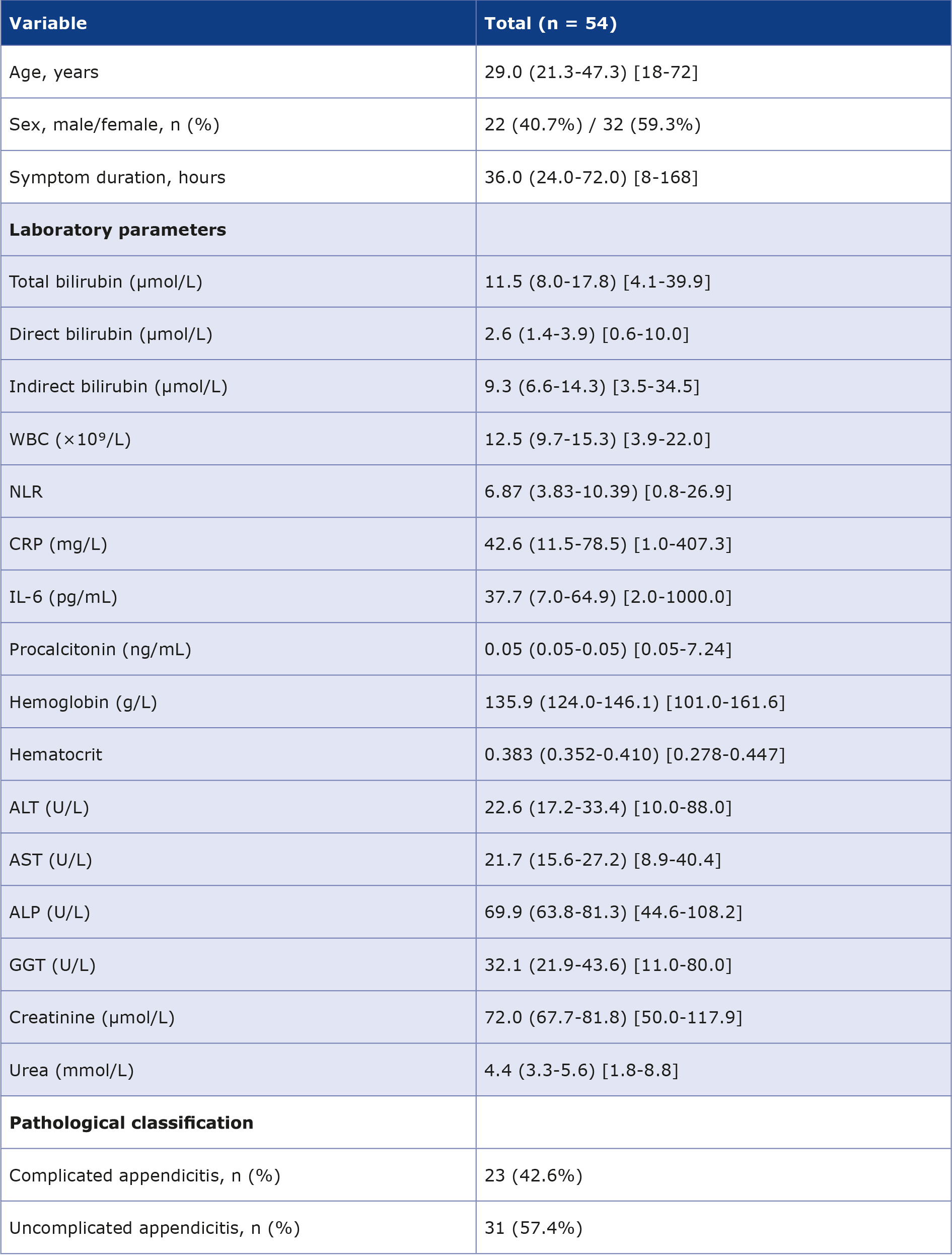
A total of 54 patients with histopathologically confirmed acute appendicitis were included in the study. 23 (42.6%) of them had complicated appendicitis (CApp) and 31 (57.4%) had uncomplicated appendicitis (non-CApp). Based on symptom duration, 19 patients (35.2%) presented within ≤ 24 hours and 35 patients (64.8%) after > 24 hours from symptom onset (Table 1).
Table 1. Baseline characteristics of the study population
Values are presented as median (interquartile range, IQR) [minimum–maximum] unless otherwise stated.
Normal ranges of laboratory parameters are presented in parentheses.
ALP – alkaline phosphatase; ALT – alanine aminotransferase; AST– aspartate aminotransferase, CRP-C–reactive protein;
GGT – gamma-glutamyl transferase; IL-6 – interleukin-6; NLR – neutrophil-to-lymphocyte ratio; WBC – white blood cell count
Total bilirubin concentrations returned to normal in all cases and remained within the laboratory reference range at follow-up, thus excluding previously unrecognized hereditary hyperbilirubinemia or persistent hepatobiliary dysfunction as causes of the pre-operative bilirubin elevations.
Symptom duration vs. laboratory parameters
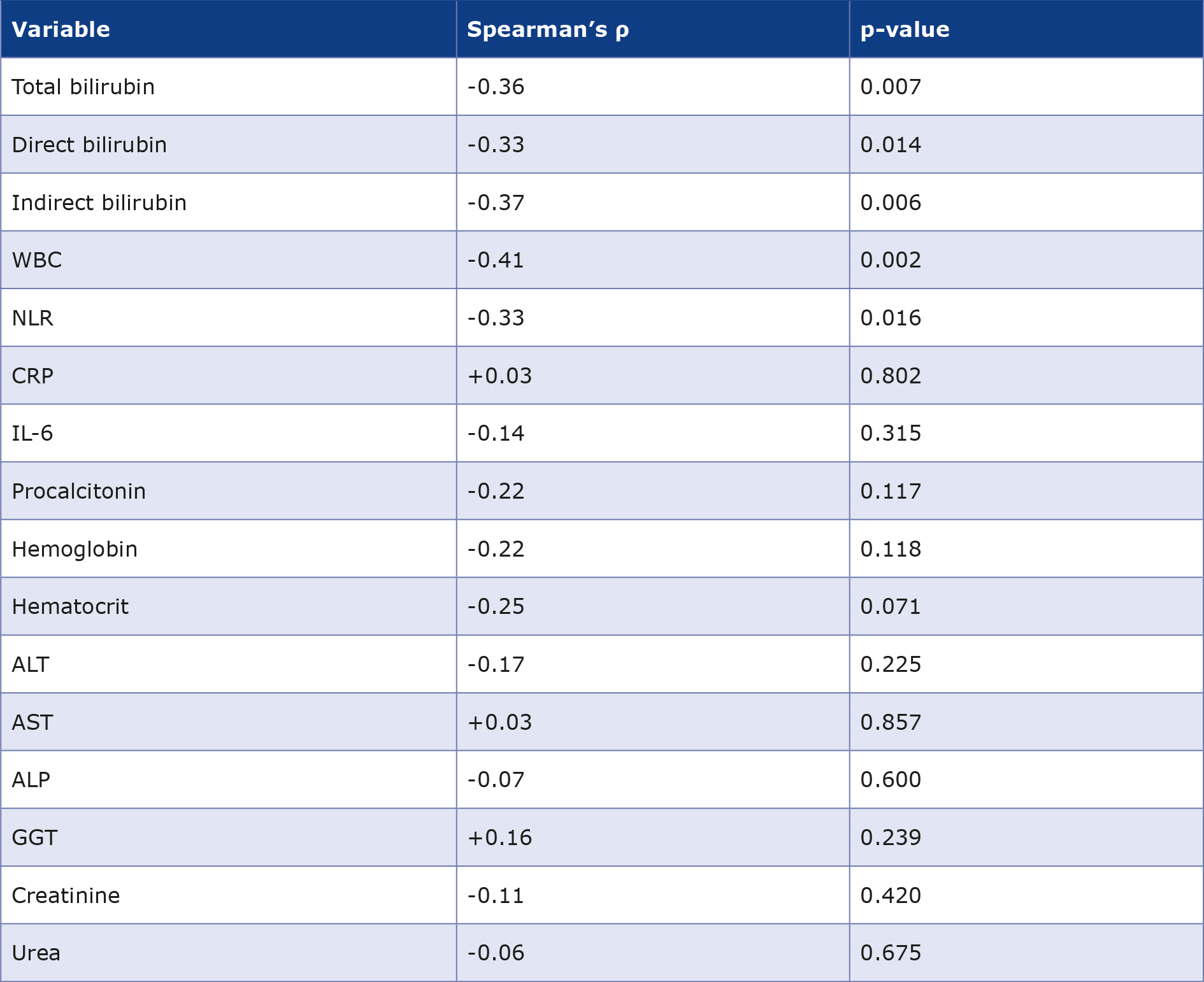
Spearman correlation analysis demonstrated significant inverse associations between symptom duration and all serum bilirubin fractions. Symptom duration correlated negatively with total bilirubin (ρ = -0.36, p = 0.007), direct bilirubin (ρ = -0.33, p = 0.014), and indirect bilirubin (ρ = -0.37, p = 0.006). Among inflammatory markers, significant inverse correlations were also observed for WBC (ρ = -0.41, p = 0.002) and NLR (ρ = -0.33, p = 0.016). In contrast, CRP (ρ = 0.03, p = 0.802), IL-6 (ρ = -0.14, p = 0.315) and PCT (ρ = -0.22, p = 0.117) did not demonstrate significant associations with symptom duration.
No significant correlations were identified between symptom duration and hemoglobin, hematocrit, ALT, AST, ALP, GGT, creatinine, or urea (all p > 0.05) (Table 2, Figure 1).
Table 2. Correlation between symptom duration and laboratory parameters
Spearman’s rank correlation coefficient (ρ) was used to assess associations between symptom duration and laboratory parameters.
ALP – alkaline phosphatase; ALT – alanine aminotransferase; AST– aspartate aminotransferase; CRP-C –reactive protein;
GGT – gamma-glutamyl transferase; IL-6 – interleukin-6; NLR – neutrophil-to-lymphocyte ratio; WBC – white blood cell count
Figure 1. Symptom duration vs. serum total bilirubin in acute appendicitis
Scatter plot demonstrating the inverse association between symptom duration and serum total bilirubin concentrations.
Each point represents an individual patient. The fitted line is shown for visual representation of the overall trend.
Symptom duration was inversely correlated with total bilirubin (Spearman’s ρ = -0.36, p = 0.007).
Symptom duration (≤ 24 h vs. > 24 h)
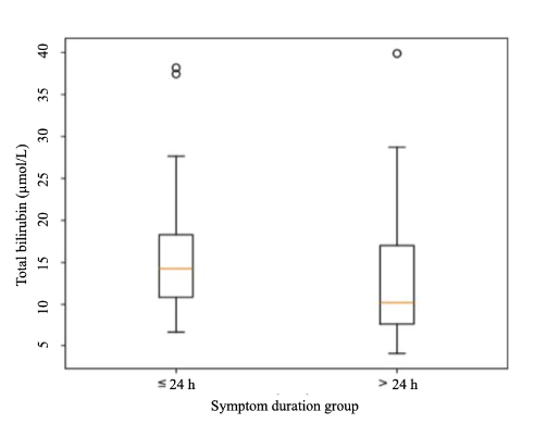
Patients presenting within ≤ 24 hours had higher concentrations of total, direct, and indirect bilirubin than patients presenting after > 24 hours. However, none of these between-group differences reached statistical significance (total bilirubin, p = 0.077; direct bilirubin, p = 0.069; indirect bilirubin, p = 0.074). Accordingly, these findings should be interpreted as descriptive and supportive rather than confirmatory. In contrast, the primary correlation analysis identified statistically significant inverse associations between symptom duration and all bilirubin fractions across the entire cohort. Among inflammatory markers, patients presenting within ≤ 24 hours had significantly higher WBC counts (p = 0.028) and NLR values (p = 0.030). No significant differences were observed for CRP, IL-6 or PCT. Similarly, no statistically significant differences were identified for hemoglobin, hematocrit, liver enzymes (ALT, AST, ALP, GGT), creatinine, or urea (all p > 0.05). These findings indicate that the observed bilirubin pattern was not accompanied by evidence of hemoconcentration, hepatocellular injury, cholestatic enzyme elevation, or renal dysfunction (Table 3, Figure 2).
Table 3. Comparison of laboratory parameters according to symptom duration (≤ 24 h vs. > 24 h)
Values are presented as median (interquartile range, IQR). Comparisons between patients presenting within ≤ 24 hours and > 24 hours from symptom onset were performed using the Mann–Whitney U test. A two-sided p-value < 0.05 was considered statistically significant.
Laboratory reference ranges: total bilirubin < 17.1 μmol/L, ALT < 52 U/L, AST < 39 U/L, ALP 46-116 U/L, GGT < 61 U/L, creatinine 53-115 μmol/L, and urea 1.7–8 mmol/L.
ALP – alkaline phosphatase; ALT – alanine aminotransferase; AST – aspartate aminotransferase; CRP-C –reactive protein; GGT – gamma-glutamyl transferase; IL-6 – interleukin-6; NLR – neutrophil-to-lymphocyte ratio; WBC – white blood cell count
Figure 2. Serum total bilirubin according to symptom duration groups
Boxplot showing serum total bilirubin concentrations in patients presenting within ≤ 24 hours and > 24 hours from symptom onset. The central line represents the median, boxes indicate the interquartile range, and whiskers represent the range. Higher bilirubin concentrations were observed in patients presenting within ≤ 24 hours, although the difference did not reach statistical significance (p = 0.077).
Analysis according to pathological severity
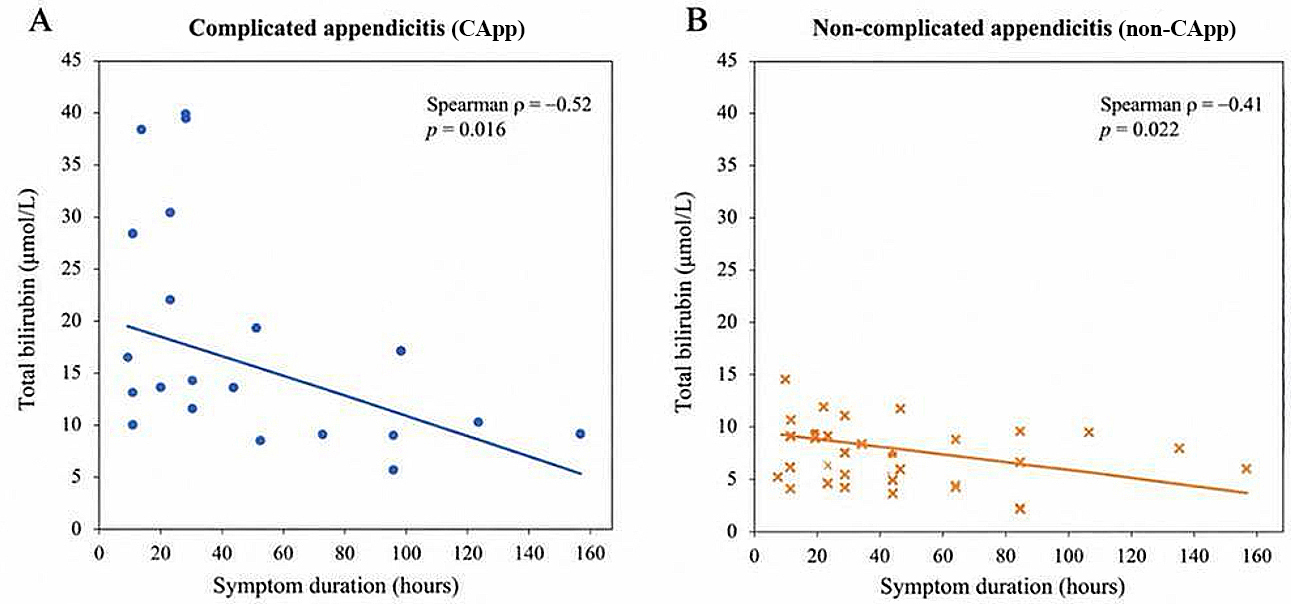
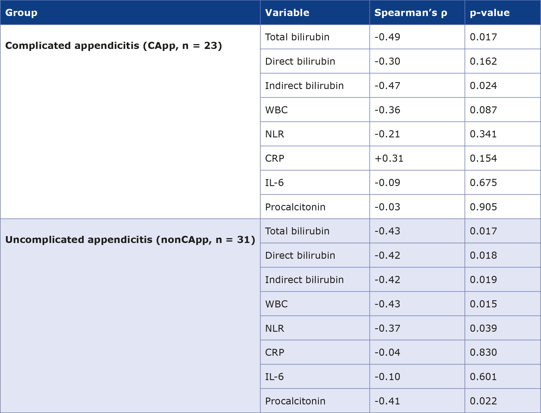
Within the CApp subgroup, symptom duration demonstrated significant inverse correlations with total bilirubin (ρ = -0.49, p = 0.017) and indirect bilirubin (ρ = -0.47, p = 0.024). Direct bilirubin showed a similar inverse trend but did not reach statistical significance (ρ = -0.30, p = 0.162). No significant associations were observed between symptom duration and WBC, NLR, CRP, IL-6 or PCT within the CApp subgroup.
Within the non-CApp subgroup, symptom duration demonstrated significant inverse correlations with all bilirubin fractions, including total bilirubin (ρ = -0.43, p = 0.017), direct bilirubin (ρ = -0.42, p = 0.018), and indirect bilirubin (ρ = -0.42, p = 0.019). Significant inverse correlations were also observed for WBC (ρ = -0.43, p = 0.015), NLR (ρ = -0.37, p = 0.039) and PCT (ρ = -0.41, p = 0.022). In contrast, CRP and IL-6 were not significantly associated with symptom duration. Collectively, a similar inverse temporal association between bilirubin and symptom duration was observed in both pathological subgroups and was not restricted to complicated appendicitis (Table 4, Figure 3).
Table 4. Correlation between symptom duration and laboratory parameters stratified by pathological severity
Values represent Spearman’s rank correlation coefficients (ρ) between symptom duration and laboratory parameters calculated separately within pathological subgroups. A two-sided p-value < 0.05 was considered statistically significant.
CRP – C-reactive protein; IL-6 – interleukin-6; NLR – neutrophil-to-lymphocyte ratio; WBC – white blood cell count
Figure 3. Symptom duration versus serum total bilirubin according to pathological severity
(A) Each point represents an individual patient. The fitted line is shown for visual representation of the overall trend. Symptom duration was inversely correlated with total bilirubin (Spearman’s ρ = -0.49, p = 0.017).
(B) Each point represents an individual patient. The fitted line is shown for visual representation of the overall trend. Symptom duration was inversely correlated with total bilirubin (Spearman’s ρ = -0.43, p = 0.017).
Discussion
The present study demonstrated significant inverse correlations between symptom duration and serum bilirubin concentrations across all bilirubin fractions. Although patients presenting within ≤ 24 hours had higher bilirubin concentrations, these subgroup differences did not reach statistical significance. Importantly, a similar temporal pattern was observed in both CApp and non-CApp, suggesting that it is not restricted to advanced pathological severity.
Our findings also reveal that different laboratory markers exhibit distinct temporal profiles. While serum bilirubin, WBC, and NLR demonstrated significant inverse associations with symptom duration, CRP, IL-6 and PCT did not show significant temporal relationships in the overall cohort. Furthermore, no significant associations were observed between symptom duration and markers of hemoconcentration (hemoglobin and hematocrit), hepatocellular injury (ALT and AST), cholestatic enzyme activity (ALP and GGT), or renal function (creatinine and urea). Collectively, our findings suggest that bilirubin may provide information regarding the host response that is not fully captured by conventional inflammatory markers alone.
Temporal dynamics of bilirubin in acute appendicitis
Previous studies have consistently demonstrated an association between hyperbilirubinemia and CApp. Jamaluddin et al., D’Souza et al., Akturk et al., Khalid and Elamin, and Basukala et al. reported that elevated bilirubin levels are associated with more severe forms of appendicitis and may assist in identifying patients at increased risk of perforation [16-20]. Similarly, Yamazaki et al. identified both bilirubin and CRP as predictors of perforated appendicitis, while Eren et al. demonstrated concurrent elevations of bilirubin, CRP, WBC and neutrophil counts in gangrenous and perforated CApp [21-22]. Adams and Jaunoo likewise reported higher bilirubin concentrations in perforated compared with non-perforated appendicitis [23].
Our study does not contradict these observations. Rather, it addresses a different aspect of bilirubin biology. Most of the previous investigations evaluated bilirubin as a static marker of disease severity, whereas our study examined its relationship with symptom duration.
The significant inverse correlations observed across the continuous range of symptom duration suggest that bilirubin concentrations vary according to the stage of appendicitis. Although dichotomization at the 24-hour threshold yielded only numerical differences, the continuous analysis supports the existence of an overall temporal association. These findings further suggest that bilirubin concentrations may increase relatively early in the disease course, including in patients who ultimately demonstrate non-CApp histopathological findings. This observation expands the traditional interpretation of hyperbilirubinemia as exclusively a marker of perforation or severe appendiceal destruction. The principal contribution of the present study therefore is characterizing the temporal behavior of bilirubin during acute appendicitis. The observed inverse relationship between bilirubin and symptom duration provides a basis for considering bilirubin within a temporal framework and suggests that biomarker interpretation may benefit from incorporating information regarding the stage of disease presentation.
Potential biological explanations
First, bacterial endotoxins and other microbial products are known to influence hepatocellular bile transport. Experimental studies have demonstrated that lipopolysaccharide can rapidly alter canalicular transporter function and impair bile excretion independently of overt structural liver injury [14, 24-27]. This may result in transient increases in serum bilirubin concentrations without corresponding elevations in conventional liver enzymes. Second, the appendix drains into the portal venous system, creating a direct anatomical pathway for delivery of microbial products to the liver. Exposure of hepatic cells to bacterial components may therefore occur early during appendiceal infection, potentially preceding widespread systemic inflammatory activation [13, 28-30]. Third, the absence of significant associations between symptom duration and ALT, AST, ALP, and GGT negates progressive hepatocellular injury or cholestatic disease as primary explanations for the observed bilirubin pattern. Similarly, the lack of significant temporal relationships for hemoglobin and hematocrit does not support hemoconcentration as the principal mechanism. Taken together, these observations support the plausibility of an early hepatobiliary response to microbial exposure. Unfortunately, direct measurement of endotoxin activity, microbial translocation and hepatocellular transport dysfunction was beyond the scope of the present study, therefore mechanistic conclusions cannot be drawn.
Bilirubin vs. inflammatory markers
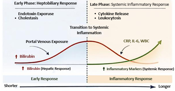
An important finding of this study is the differing temporal behavior of bilirubin and inflammatory biomarkers. WBC and NLR demonstrated inverse associations with symptom duration similar to bilirubin, whereas CRP, IL-6 and PCT did not exhibit significant temporal relationships in the overall cohort. These findings suggest that different biomarkers may reflect distinct phases or components of the host response to appendiceal inflammation. Importantly, the absence of a significant temporal relationship for CRP and IL-6 should not be interpreted as evidence that these markers lack clinical relevance. Rather, it may indicate that inflammatory biomarkers and bilirubin capture partially different biological processes and when interpreted together they may therefore provide complementary information. The partially distinct temporal profiles of bilirubin and inflammatory biomarkers form the basis of the proposed conceptual framework illustrated in Figure 4.
Figure 4. Conceptual temporal biomarker model in acute appendicitis
This hypothesized temporal framework for biomarker behavior in acute appendicitis is based on the observed cross-sectional associations. It integrates the observed inverse association between symptom duration and bilirubin concentrations with current knowledge of host–pathogen interactions and the gut–liver axis. The hypothesis is that exposure of the liver to bacterial products delivered through the portal circulation may contribute to transient elevations in serum bilirubin during earlier stages of appendicitis. As the appendicitis progresses, systemic inflammatory activation may become increasingly prominent, reflected by changes in conventional inflammatory biomarkers. This figure should not be interpreted as evidence of a validated biological sequence, causal mechanism or direct temporal progression of biomarker responses.
The “early-hit concept” as a hypothesis-generating framework
Our findings are consistent with a conceptual model in which biomarker responses in acute appendicitis might not evolve in a strictly linear fashion. Within this framework, bilirubin may represent an early component of the host response to microbial exposure, whereas other biomarkers reflect subsequent inflammatory amplification and systemic activation. Importantly, this interpretation should be viewed as hypothesis-generating rather than mechanistic. The present study is based on cross-sectional observations and cannot establish temporal causality. Consequently, Figure 4 should be interpreted as a conceptual model (intended to stimulate further investigation), rather than as a validated biological pathway.
Clinical implications
The observed inverse correlation between symptom duration and bilirubin concentrations suggests that the timing of presentation should be considered when interpreting bilirubin values. Bilirubin may provide complementary information regarding host–pathogen interactions occurring during the initial stages of appendiceal inflammation.
Conversely, the absence of bilirubin elevation in patients with longer symptom duration should not be interpreted as evidence against active disease. Consideration of symptom duration may therefore be important when interpreting bilirubin concentrations in clinical practice. Our findings do not support specific diagnostic thresholds or changes in management strategy. Rather, they highlight the potential value of incorporating temporal context into the interpretation of laboratory biomarkers.
Limitations
Key limitation of our study is its cross-sectional design which precludes direct assessment of intra-individual temporal changes and does not permit conclusions regarding causal sequencing of biomarker responses. Second, our study was conducted at a single center and included a relatively small sample size, particularly for subgroup analyses. Third, symptom duration was based on patient-reported history which is subject to recall bias. Although major confounders of bilirubin metabolism were actively excluded and postoperative normalization of bilirubin was documented in all patients, residual confounding cannot be completely excluded. Finally, direct measurements of endotoxin activity, bacterial translocation, cytokine kinetics, and hepatobiliary transporter function were not available, limiting mechanistic interpretation.
Future directions
Future prospective studies incorporating serial biomarker measurements, microbiological characterization, and direct assessment of host–pathogen interactions are required to validate the proposed temporal framework. Particular attention should be directed toward evaluating bilirubin dynamics in relation to endotoxin exposure, inflammatory signaling pathways and clinical outcomes. Such investigations may help determine whether temporal biomarker profiling can improve understanding of disease biology and contribute to more refined risk stratification in acute appendicitis.
Conclusion
Serum bilirubin demonstrated significant inverse correlations with symptom duration, indicating that higher bilirubin concentrations were associated with shorter symptom duration across the study cohort. The observed temporal relationship was generally preserved in both complicated and uncomplicated appendicitis. These findings suggest that the temporal behavior of bilirubin deserves consideration alongside pathological severity and support further investigation of bilirubin within a temporal framework. We suggest an “early-hit” concept that should be regarded as a hypothesis-generating model that requires validation through prospective studies incorporating serial biomarker measurements and direct assessment of host–pathogen interactions.
Funding
No external funding was received for this research.
Conflict of interest
The authors declare no conflicts of interest.
References
| 1. |
Di Saverio S, Podda M, De Simone B, Ceresoli M, Augustin G, Gori A, et al. Diagnosis and treatment of acute appendicitis: 2020 update of the WSES Jerusalem guidelines. World J Emerg Surg [Internet]. 2020 Dec 15;15(1):27. Available from:https://wjes.biomedcentral.com/articles/10.1186/s13017-020-00306-3
|
| 2. |
Humes DJ, Simpson J. Acute appendicitis. BMJ [Internet]. 2006;333(7567):530–4. Available from: https://www.bmj.com/lookup/doi/10.1136/bmj.38940.664363.AE
|
| 3. |
Arredondo Montero J, Bardají Pascual C, Bronte Anaut M, López-Andrés N, Antona G, Martín-Calvo N. Diagnostic performance of serum interleukin-6 in pediatric acute appendicitis: a systematic review. World J Pediatr [Internet]. 2022;18(2):91–9. Available from: https://link.springer.com/10.1007/s12519-021-00488-z
|
| 4. |
Fawkner-Corbett D, Hayward G, Alkhmees M, Van Den Bruel A, Ordóñez-Mena JM, Holtman GA. Diagnostic accuracy of blood tests of inflammation in paediatric appendicitis: a systematic review and meta-analysis. BMJ Open [Internet]. 2022;12(11):e056854. Available from: https://bmjopen.bmj.com/lookup/doi/10.1136/bmjopen-2021-056854
|
| 5. |
Yu C-W, Juan L-I, Wu M-H, Shen C-J, Wu J-Y, Lee C-C. Systematic review and meta-analysis of the diagnostic accuracy of procalcitonin, C-reactive protein and white blood cell count for suspected acute appendicitis. J Br Surg [Internet]. 2013;100(3):322–9. Available from: https://academic.oup.com/bjs/article/100/3/322/6138472
|
| 6. |
Sand M, Bechara FG, Holland-Letz T, Sand D, Mehnert G, Mann B. Diagnostic value of hyperbilirubinemia as a predictive factor for appendiceal perforation in acute appendicitis. Am J Surg [Internet]. 2009;198(2):193–8. Available from: https://linkinghub.elsevier.com/retrieve/pii/S0002961009000105
|
| 7. |
Sardoo HM, Rashidi FSM, Kohan BF, Dehdashti N, Shokoufamanesh A, Raesi R, et al. ACase-control Study on the Correlation between Serum Bilirubin Levels and Various Types of Appendicitis. Open Public Health J [Internet]. 2024;17(1). Available from: https://openpublichealthjournal.com/VOLUME/17/ELOCATOR/e18749445326476/
|
| 8. |
Gavriilidis P, De’Angelis N, Evans J, Di Saverio S, Kang P. Hyperbilirubinemia as a Predictor of Appendiceal Perforation: A Systematic Review and Diagnostic Test Meta-Analysis. J Clin Med Res [Internet]. 2019;11(3):171–8. Available from:http://www.jocmr.org/index.php/JOCMR/article/view/3724
|
| 9. |
Giordano S, Pääkkönen M, Salminen P, Grönroos JM. Elevated serum bilirubin in assessing the likelihood of perforation in acute appendicitis: A diagnostic meta-analysis. Int J Surg [Internet]. 2013;11(9):795–800. Available from: https://linkinghub.elsevier.com/retrieve/pii/S174391911300143X
|
| 10. |
Sproston NR, Ashworth JJ. Role of C-Reactive Protein at Sites of Inflammation and Infection. Front Immunol [Internet]. 2018;9. Available from: http://journal.frontiersin.org/article/10.3389/fimmu.2018.00754/full
|
| 11. |
Weidhase L, Wellhöfer D, Schulze G, Kaiser T, Drogies T, Wurst U, et al. Is Interleukin-6 abetter predictor of successful antibiotic therapy than procalcitonin and C-reactive protein? A single center study in critically ill adults. BMC Infect Dis [Internet]. 2019;19(1):150. Available from: https://bmcinfectdis.biomedcentral.com/articles/10.1186/s12879-019-3800-2
|
| 12. |
Zosimas D, Lykoudis P, Strano G, Burke J, Al-cerhan E, Shatkar V. Bilirubin is a specific marker for the diagnosis of acute appendicitis. Exp Ther Med [Internet]. 2021;22(4):1056. Available from: http://www.spandidos-publications.com/10.3892/etm.2021.10490
|
| 13. |
Brandl K, Kumar V, Eckmann L. Gut-liver axis at the frontier of host-microbial interactions. Am J Physiol Liver Physiol [Internet]. 2017;312(5):G413–9. Available from: https://www.physiology.org/doi/10.1152/ajpgi.00361.2016
|
| 14. |
Zheng Z, Wang B. The Gut-Liver Axis in Health and Disease: The Role of Gut Microbiota-Derived Signals in Liver Injury and Regeneration. Front Immunol [Internet]. 2021;12. Available from: https://www.frontiersin.org/articles/10.3389/fim-mu.2021.775526/full
|
| 15. |
Pierrakos C, Vincent J-L. Sepsis biomarkers: a review. Crit Care [Internet]. 2010;14(1):R15. Available from: https://ccforum.biomedcentral.com/articles/10.1186/cc8872
|
| 16. |
Jamaluddin M, Hussain SMA, Ahmad H. Hyperbilirubinaemia a predictive factor for complicated acute appendicitis: a study in a tertiary care hospital. J Pak Med Assoc [Internet]. 2013;63(11):1374–8. Available from: http://www.ncbi.nlm.nih.gov/pubmed/24392522
|
| 17. |
D’Souza N, Karim D, Sunthareswaran R. Bilirubin; adiagnostic marker for appendicitis. Int J Surg [Internet]. 2013;11(10):1114–7. Available from: https://linkinghub.elsevier.com/retrieve/pii/S1743919113010546
|
| 18. |
Akturk OM, Yıldırım D, Çakır M, Vardar YM. Elevated serum bilirubin levels may predict perforation of the appendix. Ann Ital Chir [Internet]. 2019;90:427–31. Available from: http://www.ncbi.nlm.nih.gov/pubmed/31814597
|
| 19. |
Khalid SY, Elamin A. The Diagnostic Accuracy of Hyperbilirubinemia in Predicting Appendicitis and Appendiceal Perforation. Cureus [Internet]. 2023; Available from: https://www.cureus.com/articles/201198-the-diagnostic-accuracy-of-hyperbiliru-binemia-in-predicting-appendicitis-and-appendiceal-perforation
|
| 20. |
Basukala S, Gurung S, Tamang A, Shrestha O, Devkota M, Thapa N, et al. Diagnostic value of biochemical markers in prediction of perforation in acute appendicitis: a cross-sectional study. Ann Med Surg [Internet]. 2024;86(11):6495–501. Available from: https://journals.lww.com/10.1097/MS9.0000000000002547
|
| 21. |
Yamazaki S, Shimodaira Y, Kobayashi A, Takata M, Hayashibara K, Sakon M, et al. Predictive factors of perforated appendicitis: Impact of the C-reactive protein level. Surg Open Sci [Internet]. 2021;6:1–4. Available from: https://linkinghub.elsevier.com/retrieve/pii/S2589845021000117
|
| 22. |
Eren T, Tombalak E, Ozemir IA, Leblebici M, Ziyade S, Ekinci O, et al. Hyperbilirubinemia as apredictive factor in acute appendicitis. Eur J Trauma Emerg Surg [Internet]. 2016;42(4):471–6. Available from: http://link.springer.com/10.1007/s00068-015-0562-4
|
| 23. |
Adams HL, Jaunoo SS. Hyperbilirubinaemia in appendicitis: the diagnostic value for prediction of appendicitis and appendiceal perforation. Eur J Trauma Emerg Surg [Internet]. 2016;42(2):249–52. Available from:http://link.springer.com/10.1007/s00068-015-0540-x
|
| 24. |
Green R, Beier D, Gollan J. Regulation of hepatocyte bile salt transporters by endotoxin and inflammatory cytokines in rodents. Gastroenterology [Internet]. 1996;111(1):193–8. Available from: https://linkinghub.elsevier.com/retrieve/pii/S0016508596003174
|
| 25. |
Trauner M, Meier PJ, Boyer JL. Molecular Pathogenesis of Cholestasis. Epstein FH, editor. N Engl J Med [Internet]. 1998 Oct 22;339(17):1217–27. Available from: http://www.nejm.org/doi/10.1056/NEJM199810223391707
|
| 26. |
Tilg H, Adolph TE, Trauner M. Gut-liver axis: Pathophysiological concepts and clinical implications. Cell Metab [Internet]. 2022;34(11):1700–18. Available from: https://linkinghub.elsevier.com/retrieve/pii/S1550413122004077
|
| 27. |
Nakadate K, Saitoh H, Sakaguchi M, Miruno F, Muramatsu N, Ito N, et al. Advances in Understanding Lipopolysaccharide-Mediated Hepatitis: Mechanisms and Pathological Features. Curr Issues Mol Biol [Internet]. 2025;47(2):79. Available from: https://www.mdpi.com/1467-3045/47/2/79
|
| 28. |
Yao Z, Mates JM, Cheplowitz AM, Hammer LP, Maiseyeu A, Phillips GS, et al. Blood-Borne Lipopolysaccharide Is Rapidly Eliminated by Liver Sinusoidal Endothelial Cells via High-Density Lipoprotein. J Immunol [Internet]. 2016;197(6):2390–9. Available from: https://academic.oup.com/jimmunol/article/197/6/2390/7964401
|
| 29. |
Topchiy E, Cirstea M, Kong HJ, Boyd JH, Wang Y, Russell JA, et al. Lipopolysaccharide Is Cleared from the Circulation by Hepatocytes via the Low Density Lipoprotein Receptor. Tancevski I, editor. PLoS One [Internet]. 2016 May 12;11(5):e0155030. Available from: https://dx.plos.org/10.1371/journal.pone.0155030
|
| 30. |
Emmanuel A, Murchan P, Wilson I, Balfe P. The value of hyperbilirubinaemia in the diagnosis of acute appendicitis. Tancevski I, editor. Ann R Coll Surg Engl [Internet]. 2011 Apr 12;93(3):213–7. Available from: https://dx.plos.org/10.1371/journal.pone.0155030
|