



Intimal and medial intracranial arterial calcifications: pathophysiology, risk factors and clinical relevance in stroke – narrative review

 , Jacek Gozdalski2, Karol Chojnowski3, Kamil Sztramski4, Małgorzata Lewalska1, Weronika Piątkowska5, Maksim Jagodziński6, Aleksandra Dańczyszyn2, Natalia Jasińska1



Abstract
Background
Intracranial arterial calcifications (ICAC) are common findings in computed tomography of stroke patients. They comprise 2 distinct entities: intimal arterial calcifications (IAC, associated with atherosclerosis) and medial arterial calcifications (MAC, related to arterial stiffening). Differentiation between these subtypes may have prognostic and therapeutic relevance.
Material and methods
We searched the PubMed, Scopus and Google Scholar databases for studies published between 1995 and 2023. Articles addressing pathophysiology, imaging characteristics, risk factors, ethnic distribution, scoring methods and clinical outcomes of ICAC in adult stroke populations were included in this narrative review.
Results
IAC appear as eccentric, irregular calcifications and are strongly linked to atherosclerotic plaques and thrombotic risk. MAC present as concentric, continuous calcifications reflecting medial degeneration and increased arterial stiffness, often promoting collateral formation. ICAC are highly prevalent in the elderly and stroke patient populations. Some studies suggest better treatment response in patients with predominant MAC, although the findings remain inconsistent.
Conclusions
IAC and MAC differ in pathophysiology, risk profiles, and potential clinical implications. Standardised differentiation may improve risk stratification and individualised stroke management.
Citation
Andruszkiewicz K, Gozdalski J, Chojnowski K, Sztramski K, Lewalska M, Piątkowska W, Jagodziński M, Dańczyszyn A, Jasińska N. Intimal and medial intracranial arterial calcifications: pathophysiology, risk factors and clinical relevance in stroke – narrative review. Eur J Transl Clin Med.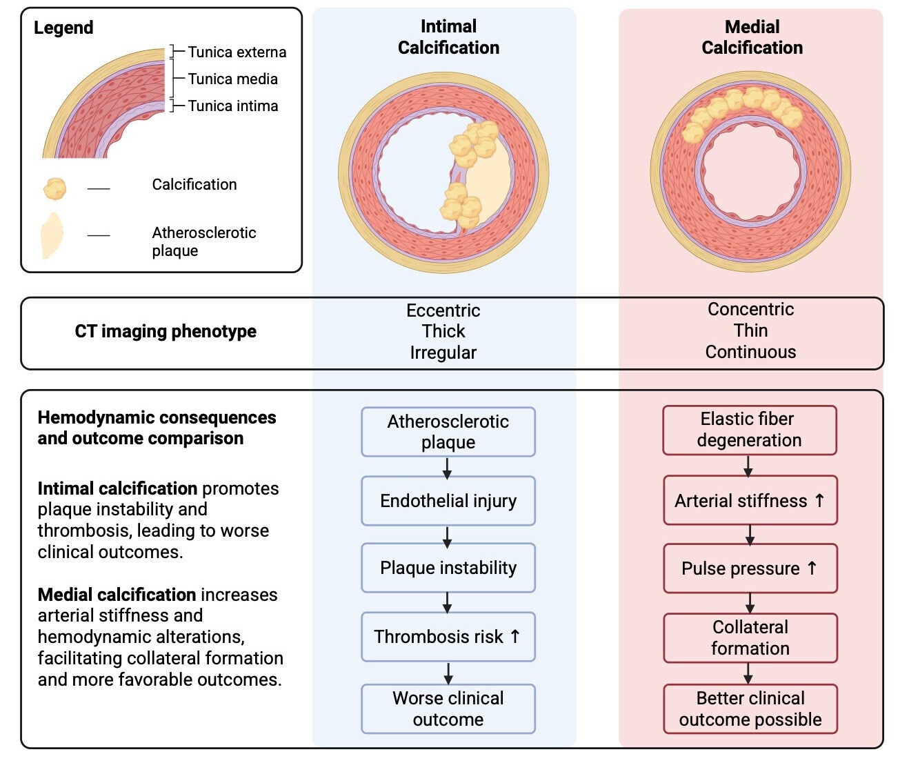


Abbreviations
- CT – computed tomography
- CTA – computed tomography angiography
- HT – hemorrhagic transformation
- IAC – intimal arterial calcifications
- ICA – internal carotid artery
- ICAC – intracranial arterial calcifications
- IEL – internal elastic lamina
- IVT – intravenous thrombolysis
- MAC – medial arterial calcifications
- mRS – modified Rankin Scale
- NIHSS – National Institute of Health Stroke Scale
Introduction
Intracranial arterial calcifications (ICAC) are frequently detected on computed tomography (CT) of stroke patients, yet their biological and clinical significance remains unclear [1]. There is increasing evidence that calcifications are not a uniform entity but can be divided into intimal and medial subtypes, which differ in pathophysiology, risk factors, hemodynamic effects and clinical outcomes [2-3]. Unfortunately, many studies still analyze ICAC as a single phenomenon, thus masking the prognostic differences [4]. This narrative review summarizes the mechanisms, classification, epidemiology, assessment, and clinical relevance of ICAC in stroke.
Material and methods
We conducted a narrative literature review to summarize the evidence on arterial calcifications and their clinical relevance in stroke. We searched the PubMed, Scopus, and Google Scholar databases for articles published in English language between 1995 and 2023. The following keywords were used: “intracranial arterial calcification”, “intimal calcification”, “medial calcification”, “Monckeberg sclerosis”, “internal elastic lamina calcification”, “stroke”, “risk factors”, “Agatston score”, “Woodcock score”, and “Kockelkoren score”. Articles were discussed by all authors and assigned to one or more sections.
No summary of appraisal of the articles was prepared, but notes were added in the text if articles contradicted each other or had different levels of evidence. Original articles addressing pathophysiology, risk factors, imaging assessment and clinical outcomes of intracranial calcifications in adult populations were included. Case reports, editorials, conference abstracts and studies unrelated to intracranial vessels were excluded. Additional articles were included in the final review after the authors’ suggestions.
Results
The PubMed search yielded 49 results, whereas in Scopus we found 57. After removal of 42 duplicates we had a total of 64 full-text articles. Google Scholar was used to find articles connected to those found in PubMed and Scopus (and within the scope of this review). This way we found 30 additional articles. We excluded a total of 51 articles and 43 articles were included in the final review.
Classification
In this review, we divided ICAC into 2 groups: intimal arterial calcifications (IAC, calcifications in the tunica intima) and medial (Monckeberg’s) calcifications in the tunica media. Some authors distinguish internal elastic lamina (IEL) calcification as a separate type [2, 5]. Due to similar etiology and hemodynamic consequences to medial calcifications, in this review we considered medial calcifications and IEL as a single group: medial arterial calcifications (MAC) [3].
Both types of calcifications differ not only in pathophysiology but also in their prevalence, localization, risk factors and influence on hemodynamics. IAC are more frequently (79.8%) associated with atherosclerotic plaques than MAC [4]. The IAC-associated plaques are eccentric and associated with luminal stenosis and intraplaque hemorrhage [4]. IAC is also highly clustered and thick in CT imaging [3]. Although MAC are considered independent of atherosclerosis, about 64.5% coexist with atherosclerotic plaques and have a strong predilection for lower limb arteries [4, 6]. In CT imaging, they present as circular, thin, continuous patterns, distinguishing them from IAC [3]. On ultrasonography calcifications appear as hyper-echoic spots or arches, typically with abluminal acoustic shadowing [6] (Figure 1).
Figure 1. Intimal and medial intracranial arterial calcification
↑ – increase, ↓ – decrease, CT – computed tomography
In the elderly population the prevalence of ICAC is 82%, they were more common in patients who suffered ischemic stroke (76-82%) than in non-ischemic stroke patients [7-9]. The most common locations of intracranial vascular calcifications include the internal carotid artery (ICA, 60-72.9% of all cases), followed by the vertebral artery (20%), middle cerebral artery (5%), and basilar artery (5%) [8-9]. The degree of ICA calcification varies, as the cavernous and supraclinoid to ophthalmic segments appear to be most affected (respectively 37.9% and 34.2%) [4]. Although IAC are generally more prevalent in intracranial arteries (61.5% of ICAC are predominantly in the ICA) [4], they are less common in the walls of intracranial part of ICA (30.9% to 43.7% of lesions), whereas MAC account for 46.9% to 56.3% [5, 10-11], while accounting for 38.5% of overall intracranial calcification. Regarding ICA, both calcification types affect predominantly its cavernous (IAC 34.3%, MAC 43.5% of ICAC) and supraclinoid to ophthalmic part (IAC 30.3%, MAC 40.3%) [4].
Both types of ICAC have different effects on hemodynamics. While current stroke management is not directly determined by ICAC subtype, differentiation between these patterns may contribute to differences in prognosis and risk stratification. Stroke treatment should focus on the patient’s underlying risk factors and ICAC type may be considered as supporting evidence to target those factors. IAC are associated mostly with atherosclerotic lesions, but their role in plaque stability is unclear [12-13]. Some authors claim that the endothelium of ICA affected predominantly by IAC may be more prone to damage, thus more often resulting in plaque disruption and thrombosis [1]. MAC influences intracranial circulation mostly via impairment of elastic fibres within arteries, causing arterial stiffening, leading to lowered compliance, distensibility, higher pulse pressure and higher pulse wave velocity [2-3, 12, 14]. These changes impair distal microcirculation and promote collateral formation [10, 15]. The presence of collateral pathways in patients with MAC may be the main reason why this group of patients has better treatment outcomes [10, 15] (Figure 1).
Epidemiology and ethnic distribution
Ethnicity is not a confirmed and independent risk factor for ICAC, although population-based differences in its prevalence and distribution have been reported. Current evidence suggests that ICAC and intracranial atherosclerosis are more frequently observed in Asian, Black, and Hispanic populations than in White populations of European descent. Sacco et al. demonstrated that Hispanic and Black populations had a higher frequency of strokes related to atherosclerotic changes in cerebral vessels [16]. The authors also noted a higher prevalence of diabetes and hypercholesterolemia in these groups.
Similar observations were reported by Gorelick et al., who found the highest prevalence of ICAC in the United States among Asian stroke patients, followed by Hispanic and Black populations, whereas White American populations showed the lowest prevalence [17]. These findings are consistent with data from the Northern Manhattan Stroke Study [16]. Fote et al. also reported increased ICAC prevalence in African, Asian, and Hispanic populations compared with White populations [18]. Several population-based studies further support these observations. Chen et al. studied 490 patients of various ages and nearly 70% of Asian participants demonstrated a significant ICAC burden [19]. In contrast, Olatunji et al. examined 130 predominantly elderly African participants and found ICAC in nearly all individuals included in the study [20]. Overall, the frequency of ICAC across ethnic groups remains insufficiently investigated because the available studies differ substantially in patient selection and methodology.
The intracranial segment of the internal carotid artery (ICA) is the most common location of calcification in all studied populations, accounting for approximately 80% of cases. Bos et al. demonstrated a high prevalence of ICAC in an elderly White European population from Rotterdam, affecting almost all of the nearly 3000 examined participants [7]. However, studies comparing populations suggest that the burden of intracranial ICA calcification may be highest in African populations, intermediate in Asian populations, and lowest in White populations [9-10, 21].
Although ethnic differences in ICAC prevalence have been observed, the underlying causes remain unclear. Variations between populations are likely influenced not only by genetic background but also by socioeconomic conditions, healthcare access and the prevalence of vascular risk factors. Further research is necessary to better understand the relationship between ethnicity and intracranial arterial calcification.
Risk factors
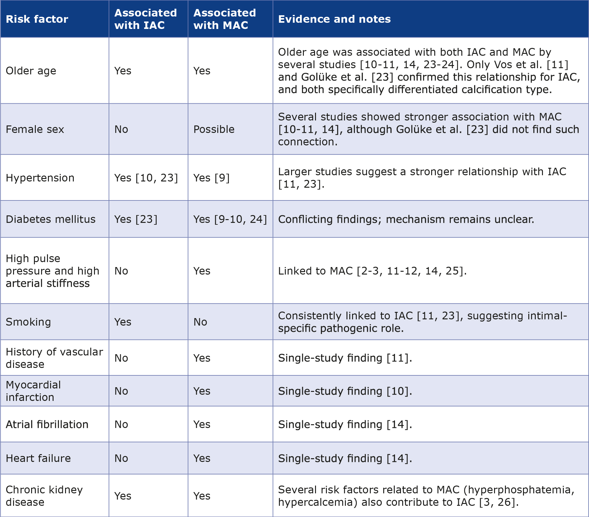
Several factors listed in Table 1 (e.g. diabetes, older age, hypertension and female sex) had inconsistent associations. Tobacco smoking seems to have a more substantial impact on IAC. Meanwhile, variables such as history of vascular diseases, myocardial infarction, atrial fibrillation and heart failure appear to be more specific to MAC. These findings suggest partially overlapping but distinct risk profiles, with different underlying mechanisms driving calcification in these arterial layers.
Table 1. Risk factors for IAC and MAC. Calcification of the intima of arteries is strongly associated with atherosclerosis, whereas calcification of media with arterial stiffness and higher pulse pressure [26].
Scoring methods
CT-based scoring methods help assess calcifications and the most widely used are the Agatston, Woodcock, modified Woodcock, and Kockelkoren scores. The Agatston score is a software-calculated calcification score weighted by the peak density of each plaque and can also deliver a calcium volume measured in cubic millimeters [22]. Although developed to measure the calcium contents in coronary arteries, it is also capable of calculating ICAC. A higher Agatston score based on total calcium score correlates with more severe calcification of the artery and higher risk for a major adverse cardiac event [23].
Woodcock and modified Woodcock scores are visual scoring methods. The Woodcock score, which considers ICA syphon calcification, determines the closest-fitting category based on the severity of the lesion [23-24]. The Woodcock method has demonstrated strong inter- and intra-rater agreement and is simpler and faster than some of the previously described visual methods [21, 23, 25-34].
Modified Woodcock Visual scoring allows more precise evaluation of calcification burden in each ICA without sacrificing speed or practicability. A number is assigned to the severity of the calcification on each axial CT, ranging from 0 to 3 [23]. Although the modified Woodcock visual score correlated strongly with the Agatston score, the study by XinWei He et al. showed a lack of correlation between the ICA modified Woodcock calcification score and the prognosis of patients with acute ischemic stroke after intravenous thrombolysis [35].
The Kockelkoren score differs from those mentioned above, as it is a tool useful in distinguishing between intimal and medial calcification dominance in the intracranial ICA [36]. Scoring was not automatic but based on the raters’ ability to detect and grade calcifications. Interrater reliability and agreement for the overall score were 0,72 (Cl: 0,60- 0,84) and 0,82 (Cl: 0,73-0,89) [3]. Scoring was performed by raters with 2 and 3 years of experience in reading CT. It should be noted that small or subtle lesions frequently produce “indistinguishable” patterns and remain “unclassified”. Calcifications were scored as “indistinguishable” mainly when presented as scarce dots of calcification, whose morphology cannot be assessed reliably [37].
Subedi et al. reported that semiautomated and volume-based scoring methods (e.g. the Agatston score) are more reliable for stroke prognosis than the simpler visual scoring methods (e.g. the Woodcock score) [23]. He et al. demonstrated that the modified Woodcock calcification score lacks a meaningful correlation with stroke outcomes, limiting its clinical utility [35]. In comparison, the Agatston and Kockelkoren scores showed promising results in risk assessment accuracy, with the latter being able to distinguish between calcification patterns.
Advanced imaging methods such as high-resolution vessel wall imaging in (HR-VWI) MRI or computed tomography angiography (CTA), may help in distinguishing calcification subtypes by enabling more precise visualisation of ICA and their walls. HR-VWI allows for image acquisition at submillimeter spatial resolution which improves diagnostic precision [38]. CTA is older than HR-VWI, but several scoring methods have been developed to assess calcifications: visual scoring, semiautomatic volume measurements and more recently, the deep-learning U-net approach (although it was tested on coronary arteries only) [35, 39].
Risk of stroke and recurrent stroke
Some authors report that higher calcification volume increases the risk of stroke [5, 40-41]. Bos et al. reported that a larger volume of ICAC correlated with a larger amount of white matter lesions in CT and a higher risk of brain infarct [42]. A similar connection was found in the Rotterdam study, whose authors claimed that ICAC may contribute to ~75% of ischemic strokes [43]. An association was also found between ICAC and the type of ischemic stroke: higher calcium volume in ICA was correlated mostly with lacunar infarcts [40].
A meta-analysis of 8 longitudinal studies involving 7297 adult patients undergoing CT or CTA demonstrated that the presence of ICAC was significantly associated with an increased risk of stroke incidence or recurrence (OR = 1.54; 95% CI 1.06-2.24) [45]. Moreover, a higher calcium burden was also positively correlated with stroke risk (OR = 1.31; 95% CI 1.17-1.46). These findings suggest that both the presence and severity of ICAC may serve as important prognostic markers for stroke risk [44]. Data on recurrent ischemic stroke in relation to calcifications is limited. Wu et al. estimated the risk of stroke recurrence as 22.5% [2]. Similar results were achieved in a cohort study done by Ovesen et al., where stenosis of 30% or greater was an independent risk factor for recurrent ischemic event [45].
Larger vascular calcification volume was related to a higher risk of post-stroke mortality and increased risk of hemorrhagic transformation after thrombolysis [2]. Authors also stated that a higher number of calcified vessels was associated with poor prognosis [12]. In patients with small-vessel occlusive stroke, the IAC Agatston score was found to be associated with a higher risk of stroke recurrence [2]. Some authors claim that there is no correlation between overall intracranial vascular calcification burden and risk of stroke. De Weert et al. compared the calcification volume in the ICAs ipsilateral to the symptomatic and asymptomatic hemispheres, however, they found no significant difference [46]. Babiarz et al. did not show any statistically significant correlation between calcification of the cavernous part of ICA and infarcts [47].
Stroke severity
Pektezel et al. reported that intracranial ICAC is particularly frequent among patients with intracerebral hematoma due to hemorrhagic stroke [14]. They also suggested that MAC may be associated with acute hematoma expansion, hospital mortality and poor prognosis. The mortality rate was higher in intracerebral hematoma patients with contralateral MAC in comparison to those with IAC and no ICAC. Nevertheless, it remains unclear whether ICAC is a direct predictor of a worse outcome. Although in univariate analysis the presence of MAC in both ipsilateral and contralateral sides was associated with poorer prognosis, in different logistic modeling only 2 parameters measured upon admission (the volume of hematoma and NIHSS) were found to be negative predictors of prognosis.
Yu et al. concluded that the increase in calcification volume on the ipsilateral side results in a significant increase in the risk of hemorrhagic transformation [12]. Moreover, the risk of poor prognosis after 3 months is increased for each additional number of calcified vessels. NIHSS, calcification volume on the ipsilateral side, overall calcification volume and the number of calcified vessels, were all significantly higher in the hemorrhagic transformation group than in the rest of the patients in the study.
A meta-analysis conducted in 2025 showed an association between ICAC and poor functional outcome at 3 months [48]. Nevertheless, the data from the included studies were suboptimal, with a variation in the reporting of outcome measures and follow-up of subjects [48]. Wu et al. found that ICAC is a meaningful risk factor for recurrent stroke, and poststroke mortality [2]. Patients with higher Agatston score had an increased risk of recurrent stroke independent of cardiovascular risk factors and independently related to a higher risk of post-stroke mortality.
Impact on cognitive function and outcomes
Studies suggest that ICAC is associated not only with a higher risk of stroke and worse functional outcomes after treatment, but also with a higher risk of cognitive decline and dementia. Bos et al. found that larger CT-assessed calcification volume is related to smaller white matter volumes and worse microstructural integrity, which may explain poorer cognitive performance in the studied population [49]. In their prospective cohort study, Seyman et al. used a study-specific semiquantitative intracranial calcification score (unlike the Agatston, Woodcock, modified Woodcock, or Kockelkoren scores discussed above), which independently predicted cognitive deterioration 24 months after the stroke in the studied population of mild stroke survivors with intact cognition, which may suggest the potential role of ICAC as a predictor of future post-stroke cognitive impairment [50].
Clinical outcome of treatment after acute ischemic stroke
Some researchers suggest that the specific pattern of intracranial carotid artery calcification may play a role in treatment outcomes. In a recent prospective study, intravenous thrombolysis (IVT) was associated with a significantly more favourable clinical outcome in the group without ICAC and in the group with a MAC pattern, but not in the group with an IAC or “indistinguishable” ICAC pattern [15]. In the MR CLEAN subgroup analysis, patients with predominantly medial calcification showed better functional outcome of endovascular treatment, compared to patients with IAC [10]. Several mechanisms have been suggested. Calcification of the medial layer leads to arterial stiffening and, consequently, to increasing pulse pressure, which causes impaired regulation of blood flow distally to the calcification site. Consequently, this may lead to an impaired distal microvascular cerebral perfusion and may trigger the formation of collateral vessels [10, 15]. Another reason could be the differences in the thrombus between patients with intimal and medial calcifications, which may result in less effective IVT in patients with predominantly IAC [10]. However, there are studies that showed no correlation between ICAC and prognosis after IVT in acute ischemic stroke [47].
Assessment of the secondary outcome of IVT also shows ambiguous results. One study found that the follow-up infarct volume or intracranial hemorrhage was not significant for any of the ICAC pattern and the effect on recanalization was only significant in the group with a predominantly MAC pattern [15]. HT is a major complication of intravenous thrombolysis, which can greatly deteriorate the patient’s prognosis. The correlation between intracranial calcification and HT remains unclear. A 2013 study showed that a significantly higher percentage of patients with moderate to severe ICAC developed intracerebral hemorrhage following IVT [46]. In a study from 2018, which was the first to examine the relationship between ICAC subtypes and the response to thrombolysis, patients with MAC had a higher risk of HT and worse reactivity to thrombolytic drugs compared to patients with IAC [48]. Another study suggested that the higher calcification volume on the lesion side was significantly associated with a higher risk of HT after IVT [12]. The pathophysiologic explanation of these results could be the worsening of vascular elasticity, causing damage to vascular autoregulation. In the presence of ischemic lesions, this could lead to higher hydrostatic pressure in the capillary bed, resulting in blood-brain barrier leakage and HT [12].
Beyond IVT and endovascular treatment, differentiation between intimal and medial intracranial arterial calcifications may also have implications for long-term secondary prevention after ischemic stroke. Since IAC is strongly associated with intracranial atherosclerosis, plaque burden, and thrombotic risk [1, 4, 11], patients with predominant IAC may benefit from intensive vascular risk-reduction strategies similar to those recommended in current guidelines for symptomatic intracranial atherosclerotic disease, including aggressive medical management based on antiplatelet therapy, intensive lipid lowering, strict blood pressure control and management of vascular risk factors [49]. In contrast, MAC is more closely associated with arterial stiffness, impaired vascular compliance, and altered cerebral hemodynamics, suggesting that optimising blood pressure control and preserving cerebral microvascular perfusion may be particularly relevant for these patients [3, 10, 15]. Unfortunately, current evidence remains insufficient to support calcification pattern-guided therapeutic strategies, and further prospective studies are needed to determine whether differentiation between IAC and MAC should influence individualised stroke prevention and treatment approaches.
Our review demonstrated that the relationship between ICAC and the clinical outcome of IVT and/or endovascular treatment after acute ischemic stroke has not been explored fully. Some studies suggest the pattern of ICAC may be a factor determining the primary clinical outcome, as well as the risk of HT and prognosis after IVT. Further exploration of this topic may bring valuable results and create new prognostic markers for the selection of patients for IVT or requiring stricter observation.
Conclusion
Studies indicate that ICAC are associated with a higher risk of intracranial hemorrhage, greater hematoma volume, hemorrhagic transformation after IVT, and worse clinical outcomes, suggesting that ICAC presence should be considered during acute stroke management. There is a lack of comparative studies that assess this risk in relation to patients without calcifications. Multicenter randomized or large registry-based studies would provide the statistical power necessary to determine whether calcification subtypes should guide patient selection for IVT or mechanical thrombectomy. In ethnically diverse countries, it seems reasonable to conduct research and treatment considering the increased risk in certain groups. These differences may arise from genetic and environmental factors. Future studies should account for socioeconomic confounders when assessing ethnic differences. Standardized criteria should be created for measurement of calcifications. Researchers should adhere to a unified classification distinguishing IAC from MAC, e.g. the Kockelkoren score. Complementary use of the Agatston score for quantifying overall calcification burden is recommended, as it demonstrates superior prognostic reliability compared to visual methods. Comparative studies are necessary to select the optimal method for specific clinical situations.
Funding
No external funding was received for this research.
Conflict of interest
The authors declare no conflicts of interest.
References
| 1. |
Gurel K, Khasiyev F, Spagnolo-Allende A, Rahman S, Liu M, Kulick ER, et al. The role of intracranial artery calcification (IAC) in stroke subtype and risk of vascular events. J Stroke Cerebrovasc Dis [Internet]. 2023;32(8):107185. Available from: https://www.sciencedirect.com/science/article/pii/S1052305723002082
|
| 2. |
Wu X, Bos D, Ren L, Leung TW, Chu WC-W, Wong LKS, et al. Intracranial Arterial Calcification Relates to Long-Term Risk of Recurrent Stroke and Post-stroke Mortality. Front Neurol [Internet]. 2020 Oct 9;11. Available from: https://www.frontiersin.org/article/10.3389/fneur.2020.559158/full
|
| 3. |
Kockelkoren R, Vos A, Van Hecke W, Vink A, Bleys RLAW, Verdoorn D, et al. Computed Tomographic Distinction of Intimal and Medial Calcification in the Intracranial Internal Carotid Artery. Cheng X, editor. PLoS One [Internet]. 2017 Jan 6;12(1):e0168360. Available from: https://dx.plos.org/10.1371/journal.pone.0168360
|
| 4. |
Du H, Li J, Yang W, Bos D, Zheng L, Wong LKS, et al. Intracranial Arterial Calcification and Intracranial Atherosclerosis: Close but Different. Front Neurol [Internet]. 2022;13. Available from:https://www.frontiersin.org/articles/10.3389/fneur.2022.799429/full
|
| 5. |
van den Beukel TC, van der Toorn JE, Vernooij MW, Kavousi M, Akyildiz AC, de Jong PA, et al. Morphological Subtypes of Intracranial Internal Carotid Artery Arteriosclerosis and the Risk of Stroke. Stroke [Internet]. 2022 Apr;53(4):1339–47. Available from: https://www.ahajournals.org/doi/10.1161/STROKEAHA.121.036213
|
| 6. |
Lanzer P, Schurgers L, Twarda-Clapa A, Ferraresi R, Hui H, Kamenskiy A, et al. Medial arterial calcification in ageing and disease: current evidence and knowledge gaps. Eur Heart J [Internet]. 2025;46(45):4876–900. Available from:https://academic.oup.com/eurheartj/article/46/45/4876/8176446
|
| 7. |
Bos D, van der Rijk MJM, Geeraedts TEA, Hofman A, Krestin GP, Witteman JCM, et al. Intracranial Carotid Artery Atherosclerosis. Stroke [Internet]. 2012;43(7):1878–84. Available from: https://www.ahajournals.org/doi/10.1161/STROKEA-HA.111.648667
|
| 8. |
Saade C, Najem E, Asmar K, Salman R, El Achkar B, Naffaa L. Intracranial calcifications on CT: an updated review. J Radiol Case Rep [Internet]. 2019;13(8):1–18. Available from: https://www.radiologycases.com/index.php/radiologycases/article/view/3633
|
| 9. |
Bugnicourt J-M, Chillon J-M, Massy ZA, Canaple S, Lamy C, Deramond HAA, et al. High Prevalence of Intracranial Artery Calcification in Stroke Patients with CKD. Clin J Am Soc Nephrol [Internet]. 2009;4(2):284–90. Available from:https://www.ovid.com/01277230-200902000-00007
|
| 10. |
Compagne KCJ, Clephas PRD, Majoie CBLM, Roos YBWEM, Berkhemer OA, van Oostenbrugge RJ, et al. Intracranial Carotid Artery Calcification and Effect of Endovascular Stroke Treatment. Stroke [Internet]. 2018;49(12):2961–8. Available from: https://www.ahajournals.org/doi/10.1161/STROKEAHA.118.022400
|
| 11. |
Vos A, Kockelkoren R, de Vis JB, van der Schouw YT, van der Schaaf IC, Velthuis BK, et al. Risk factors for atherosclerotic and medial arterial calcification of the intracranial internal carotid artery. Atherosclerosis [Internet]. 2018;276:44–9. Available from: https://www.sciencedirect.com/science/article/pii/S0021915018312048
|
| 12. |
Yu Y, Zhang F-L, Qu Y-M, Zhang P, Zhou H-W, Luo Y, et al. Intracranial Calcification is Predictive for Hemorrhagic Transformation and Prognosis After Intravenous Thrombolysis in Non-Cardioembolic Stroke Patients. J Atheroscler Thromb [Internet]. 2021;28(4):356–64. Available from: https://www.jstage.jst.go.jp/article/jat/28/4/28_55889/_article
|
| 13. |
Johnson RC, Leopold JA, Loscalzo J. Vascular Calcification. Circ Res [Internet]. 2006;99(10):1044–59. Available from: https://www.ahajournals.org/doi/10.1161/01.RES.0000249379.55535.21
|
| 14. |
Pektezel MY, Arsava EM, Gocmen R, Topcuoglu MA. Intracerebral hematoma expansion and intracranial internal carotid artery calcifications. Clin Neurol Neurosurg [Internet]. 2021;200:106361. Available from: https://linkinghub.elsevier.com/retrieve/pii/S0303846720307046
|
| 15. |
Kauw F, de Jong PA, Takx RAP, de Jong HWAM, Kappelle LJ, Velthuis BK, et al. Effect of intravenous thrombolysis in stroke depends on pattern of intracranial internal carotid artery calcification. Atherosclerosis [Internet]. 2021;316:8–14. Available from: https://www.sciencedirect.com/science/article/pii/S0021915020315288
|
| 16. |
Sacco RL, Kargman DE, Gu Q, Zamanillo MC. Race-Ethnicity and Determinants of Intracranial Atherosclerotic Cerebral Infarction. Stroke [Internet]. 1995;26(1):14–20. Available from: https://doi.org/10.1161/01.STR.26.1.14
|
| 17. |
Gorelick PB, Wong KS, Bae H-J, Pandey DK. Large Artery Intracranial Occlusive Disease. Stroke [Internet]. 2008;39(8):2396–9. Available from: https://www.ahajournals.org/doi/10.1161/STROKEAHA.107.505776
|
| 18. |
Fote GM, Raefsky S, Mock K, Chaudhari A, Shafie M, Yu W. Intracranial Arterial Calcifications: Potential Biomarkers of Stroke Risk and Outcome. Front Neurol [Internet]. 2022;13. Available from: https://www.frontiersin.org/articles/10.3389/fneur.2022.900579/full
|
| 19. |
Chen X, Lam WWM, Ng HK, Fan Y, Wong KS. The Frequency and Determinants of Calcification in Intracranial Arteries in Chinese Patients Who Underwent Computed Tomography Examinations. Cerebrovasc Dis [Internet]. 2006;21(1–2):91–7. Available from: https://karger.com/article/doi/10.1159/000090206
|
| 20. |
Olatunji RB, Adekanmi AJ, Ogunseyinde AO. Intracranial Arterial Calcification in Black Africans with Acute Ischaemic Stroke. Cerebrovasc Dis Extra [Internet]. 2018;8(1):26–38. Available from: https://karger.com/article/doi/10.1159/000485195
|
| 21. |
Chen X, Lam WWM, Ng HK, Fan Y, Wong KS. Intracranial Artery Calcification: A Newly Identified Risk Factor of Ischemic Stroke. J Neuroimaging [Internet]. 2007;17(4):300–3. Available from: https://onlinelibrary.wiley.com/doi/10.1111/j.1552-6569.2007.00158.x
|
| 22. |
Agatston AS, Janowitz WR, Hildner FJ, Zusmer NR, Viamonte M, Detrano R. Quantification of coronary artery calcium using ultrafast computed tomography. J Am Coll Cardiol [Internet]. 1990;15(4):827–32. Available from: https://linkinghub.elsevier.com/retrieve/pii/073510979090282T
|
| 23. |
Golüke NMS, de Brouwer EJM, de Jonghe A, Claus JJ, Staekenborg SS, Emmelot-Vonk MH, et al. Intracranial artery calcifications: Risk factors and association with cardiovascular disease and cognitive function. J Neuroradiol [Internet]. 2022;49(3):281–7. Available from: https://linkinghub.elsevier.com/retrieve/pii/S0150986120302418
|
| 24. |
Kim J-M, Park K-Y, Bae J-H, Han S-H, Jeong H-B, Jeong D. Intracranial Arterial Calcificationes Can Reflect Cerebral Atherosclerosis Burden. J Clin Neurol [Internet]. 2019;15(1):38. Available from: https://www.thejcn.com/DOIx.php?id=10.3988/jcn.2019.15.1.38
|
| 25. |
Amann K. Media Calcification and Intima Calcification Are Distinct Entities in Chronic Kidney Disease. Clin J Am Soc Nephrol [Internet]. 2008;3(6):1599–605. Available from: https://www.ovid.com/01277230-200811000-00003
|
| 26. |
Lucci C, Rissanen I, van den Beukel TC, Takx R, de Jong PA, Hendrikse J, et al. Risk Factors for Medial and Intimal Intracranial Internal Carotid Artery Calcification in Men and Women with Cardiovascular Disease: The UCC-SMART Study. Cerebrovasc Dis [Internet]. 2024;1–9. Available from: https://karger.com/article/doi/10.1159/000536422
|
| 27. |
Subedi D, Zishan US, Chappell F, Gregoriades M-L, Sudlow C, Sellar R, et al. Intracranial Carotid Calcification on Cranial Computed Tomography. Stroke [Internet]. 2015;46(9):2504–9. Available from: https://www.ahajournals.org/doi/10.1161/STROKEAHA.115.009716
|
| 28. |
Woodcock Jr RJ, Goldstein JH, Kallmes DF, Cloft HJ, Phillips CD. Angiographic Correlation of CT Calcification in the Carotid Siphon. Am J Neuroradiol [Internet]. 1999;20(3):495 LP – 499. Available from: http://www.ajnr.org/content/20/3/495.abstract
|
| 29. |
Ptak T, Hunter G, Avakian R, Novelline R. Clinical Significance of Cavernous Carotid Calcifications Encountered on Head Computed Tomography Scans Performed on Patients Seen in the Emergency Department. J Comput Assist Tomogr. 2003;27(4):505–9.
|
| 30. |
Bugnicourt J-M, Leclercq C, Chillon J-M, Diouf M, Deramond H, Canaple S, et al. Presence of Intracranial Artery Calcification Is Associated With Mortality and Vascular Events in Patients With Ischemic Stroke After Hospital Discharge. Stroke [Internet]. 2011;42(12):3447–53. Available from: https://www.ahajournals.org/doi/10.1161/STROKEAHA.111.618652
|
| 31. |
Savy LE, Moseley IF. Intracranial arterial calcification and ectasia in visual failure. Br J Radiol [Internet]. 1996;69(821):394–401. Available from: https://academic.oup.com/bjr/article/69/821/394-401/7316949
|
| 32. |
Suzuki M, Ozaki Y, Komura S, Nakanishi A. Intracranial carotid calcification on CT images as an indicator of atheromatous plaque: analysis of high-resolution CTA images using a 64-multidetector scanner. Radiat Med [Internet]. 2007;25(8):378–85. Available from: http://link.springer.com/10.1007/s11604-007-0153-3
|
| 33. |
Erbay SH, O’Callaghan M, Shah P, Kini J, Bassett Midle J, Polak JF. Prospective Evaluation of the Role of Atherosclerosis on Cerebral Atrophy: Pilot Study. J Neuroimaging [Internet]. 2008;18(4):375–80. Available from: https://onlinelibrary.wiley.com/doi/10.1111/j.1552-6569.2007.00149.x
|
| 34. |
Bleeker L, Marquering HA, van den Berg R, Nederkoorn PJ, Majoie CB. Semi-automatic quantitative measurements of intracranial internal carotid artery stenosis and calcification using CT angiography. Neuroradiology [Internet]. 2012;54(9):919–27. Available from: http://link.springer.com/10.1007/s00234-011-0998-0
|
| 35. |
He X-W, Zhao R, Li G-F, Zheng B, Wu Y-L, Shi Y-H, et al. Lack of Correlation Between Intracranial Carotid Artery Modified Woodcock Calcification Score and Prognosis of Patients With Acute Ischemic Stroke After Intravenous Thrombolysis. Front Neurol [Internet]. 2019;10. Available from: https://www.frontiersin.org/article/10.3389/fneur.2019.00696/full
|
| 36. |
Hong NR, Seo HS, Lee YH, Kim JH, Seol HY, Lee NJ, et al. The correlation between carotid siphon calcification and lacunar infarction. Neuroradiology [Internet]. 2011;53(9):643–9. Available from: http://link.springer.com/10.1007/s00234-010-0798-y
|
| 37. |
Kassab MY, Gupta R, Majid A, Farooq MU, Giles BP, Johnson MD, et al. Extent of Intra-Arterial Calcification on Head CT Is Predictive of the Degree of Intracranial Atherosclerosis on Digital Subtraction Angiography. Cerebrovasc Dis [Internet]. 2009;28(1):45–8. Available from: https://karger.com/article/doi/10.1159/000219296
|
| 38. |
Ahn SS, Nam HS, Heo JH, Kim YD, Lee S-K, Han K, et al. Quantification of intracranial internal carotid artery calcification on brain unenhanced CT: evaluation of its feasibility and assessment of the reliability of visual grading scales. Eur Radiol [Internet]. 2013;23(1):20–7. Available from: http://link.springer.com/10.1007/s00330-012-2586-z
|
| 39. |
Kockelkoren R, Jairam PM, Murchison JT, Debray TPA, Mirsadraee S, van der Graaf Y, et al. Validation of an imaging based cardiovascular risk score in a Scottish population. Eur J Radiol [Internet]. 2018;98:143–9. Available from: https://linkinghub.elsevier.com/retrieve/pii/S0720048X17305065
|
| 40. |
Zwakenberg SR, de Jong PA, Hendriks EJ, Westerink J, Spiering W, de Borst GJ, et al. Intimal and medial calcification in relation to cardiovascular risk factors. Metzinger L, editor. PLoS One [Internet]. 2020;15(7):e0235228. Available from:https://dx.plos.org/10.1371/journal.pone.0235228
|
| 41. |
Kang D-W, Kim DY, Kim J, Baik SH, Jung C, Singh N, et al. Emerging Concept of Intracranial Arterial Diseases: The Role of High Resolution Vessel Wall MRI. J Stroke [Internet]. 2024;26(1):26–40. Available from: http://j-stroke.org/journal/view.php?-doi=10.5853/jos.2023.02481
|
| 42. |
Bos D, Ikram MA, Elias-Smale SE, Krestin GP, Hofman A, Witteman JCM, et al. Calcification in Major Vessel Beds Relates to Vascular Brain Disease. Arterioscler Thromb Vasc Biol [Internet]. 2011;31(10):2331–7. Available from: https://www.ahajournals.org/doi/10.1161/ATVBAHA.111.232728
|
| 43. |
Bos D, Portegies MLP, van der Lugt A, Bos MJ, Koudstaal PJ, Hofman A, et al. Intracranial Carotid Artery Atherosclerosis and the Risk of Stroke in Whites. JAMA Neurol [Internet]. 2014;71(4):405. Available from: http://archneur.jamanetwork.com/article.aspx?doi=10.1001/jamaneurol.2013.6223
|
| 44. |
Conte M, Alalfi MO, Cau R, Scicolone R, Chaturvedi S, Virmani R, et al. Cracking the Code of Calcification: How Presence and Burden among Intracranial Arteries Influence Stroke Incidence and Recurrence. Am J Neuroradiol [Internet]. 2025;ajnr. A8668. Available from: http://www.ajnr.org/lookup/doi/10.3174/ajnr.A8668
|
| 45. |
Ovesen C, Abild A, Christensen AF, Rosenbaum S, Hansen CK, Havsteen I, et al. Prevalence and long-term clinical significance of intracranial atherosclerosis after ischaemic stroke or transient ischaemic attack: a cohort study. BMJ Open [Internet]. 2013;3(10):e003724. Available from: https://bmjopen.bmj.com/lookup/doi/10.1136/bmjopen-2013-003724
|
| 46. |
de Weert TT, Cakir H, Rozie S, Cretier S, Meijering E, Dippel DWJ, et al. Intracranial Internal Carotid Artery Calcifications: Association with Vascular Risk Factors and Ischemic Cerebrovascular Disease. Am J Neuroradiol [Internet]. 2009;30(1):177–84. Available from: http://www.ajnr.org/lookup/doi/10.3174/ajnr.A1301
|
| 47. |
Babiarz LS, Yousem DM, Bilker W, Wasserman BA. Middle Cerebral Artery Infarction: Relationship of Cavernous Carotid Artery Calcification. Am J Neuroradiol [Internet]. 2005;26(6):1505 LP – 1511. Available from: http://www.ajnr.org/con-tent/26/6/1505.abstract
|
| 48. |
Elsheikh S, McKeever S, Irving GJ, Hill AM, Lip GY, Abdul-Rahim AH. Intracranial arterial calcification as a marker of stroke risk and worse stroke outcomes in adults: A systematic review and meta-analysis. Int J Stroke [Internet]. 2026;21(3):335–42. Available from: https://journals.sagepub.com/doi/10.1177/17474930251372693
|
| 49. |
Bos D, Vernooij MW, Elias-Smale SE, Verhaaren BFJ, Vrooman HA, Hofman A, et al. Atherosclerotic calcification relates to cognitive function and to brain changes on magnetic resonance imaging. Alzheimer’s Dement [Internet]. 2012;8(5S). Available from: https://alz-journals.onlinelibrary.wiley.com/doi/10.1016/j.jalz.2012.01.008
|
| 50. |
Seyman EE, Sadeh-Gonik U, Berman P, Blum I, Shendler G, Nathan B, et al. Association between intracranial vessel calcifications, structural brain damage, and cognitive impairment after minor strokes: a prospective study. Front Neurol [Internet]. 2023;14. Available from: https://www.frontiersin.org/articles/10.3389/fneur.2023.1218077/full
|
