

Diagnostic accuracy of urinary NGAL compared to the gold standard imaging for detecting renal scarring in pediatric vesicoureteral reflux: a systematic review and meta-analysis





Abstract
Introduction: Vesicoureteral reflux (VUR) in children can cause recurrent urinary tract infections and renal scarring. Although 99mTc-DMSA scintigraphy is the reference test, it is invasive and involves ionising radiation. Urinary neutrophil gelatinase-associated lipocalin (uNGAL) may be a non-invasive marker of renal injury.
Material and methods: Following PRISMA 2020, we searched PubMed, ScienceDirect, Europe PMC, OpenAlex, Taylor & Francis Online, and Google Scholar for studies of pediatric (< 18 years of age) VUR comparing uNGAL with DMSA for scar detection. Diagnostic accuracy was pooled using a Bayesian bivariate random-effects model. Risk of bias was assessed with QUADAS-2.
Results: Four studies were included. uNGAL was reported as absolute values or creatinine-normalized (uNGAL/Cr), although meta-analysis was feasible only for uNGAL/Cr. Pooled sensitivity was 0.72 (95% CI 0.52-0.93) and specificity 0.63 (95% CI 0.15-0.85), with diagnostic odds ratio 4.18 (95% CI 1.17- 17.70). SROC indicated moderate performance with substantial heterogeneity. Overall risk of bias was moderate.
Conclusions: uNGAL/Cr shows moderate accuracy for detecting renal scarring in pediatric VUR and may complement, but not replace, DMSA to help limit radiation exposure. Larger multi-center studies with standardized cut-offs are needed.
Citation
Rachman Y, Nurhadi P, Purnomo A F. Diagnostic accuracy of urinary NGAL compared to the gold standard imaging for detecting renal scarring in pediatric vesicoureteral reflux: a systematic review and meta-analysis. Eur J Transl Clin Med.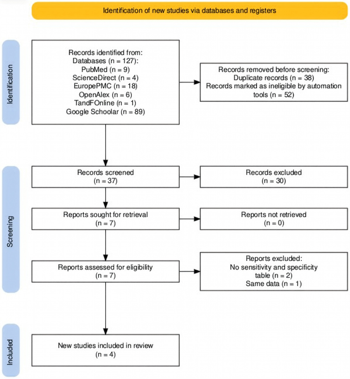








Introduction
Vesicoureteral reflux (VUR) is one of the most prevalent congenital anomalies affecting the urinary tract in children, impacting approximately 1% to 2% of the pediatric population [1-3]. This condition is characterised by the abnormal retrograde flow of urine from the bladder into the ureters and renal collecting system [1, 4]. VUR significantly increases the risk of urinary tract infections (UTIs) in children [1, 4]. The most significant long-term complication of VUR is the development of renal scarring [1-2, 4-6], traditionally termed reflux nephropathy (RN) [7]. This progressive condition can lead to severe sequelae including proteinuria, arterial hypertension, and chronic kidney disease (CKD) [7]. 7-17% of cases can become end-stage renal disease (ESRD) because of reflux nephropathy [7]. VUR is indeed a notable etiology in 8.4% of pediatric CKD cases and 6% of ESRD cases in children and young adults [2].
The definitive diagnosis of VUR typically relies on voiding cystourethrography (VCUG), while the gold standard imaging method for detecting renal scarring is 99mTc-dimercaptosuccinic acid (DMSA) renal scintigraphy [1, 4-6, 8-9]. However, both of these imaging procedures expose children to ionising radiation, are invasive, expensive and cannot be readily repeated for routine monitoring [4, 8, 10]. Given the socio-economic burden of recurrent hospitalisations and the critical need for early diagnosis and appropriate management of VUR, there is a pressing demand for novel, non-invasive, and easily quantifiable biomarkers to facilitate diagnosis, staging, long-term follow-up, and prevention of complications [1, 8, 11-12]. There are several studies actively searching for reliable, non-invasive biochemical markers that can be objectively measured to identify patients at high risk for renal damage. Previous studies reported neutrophil gelatinase-associated lipocalin (NGAL) as the biochemical marker that increased significantly in pediatric VUR patient with renal scarring condition [5-6, 9, 13].
NGAL is a 25-kDa glycoprotein belonging to the lipocalin superfamily [13-14]. NGAL is rapidly induced by various cell types, including immune cells, hepatocytes, and renal tubular cells, in response to cellular stress such as inflammation and ischemia [14-16]. It is well-established as a good diagnostic and prognostic marker for acute kidney injury (AKI) and is increasingly recognised for its potential in early identification and prediction of CKD progression [15-20]. Elevated urinary NGAL (uNGAL) concentrations are observed in kidney damage due to increased production and decreased reabsorption by renal tubular cells with this elevation potentially acting as a protective mechanism to limit kidney injury [14-15]. Furthermore, a meta-analysis has identified an optimal cut-off value of 48.43 ng/mL for uNGAL, demonstrating high sensitivity (96%) and specificity (97%) for diagnosing UTIs in children, highlighting its utility as a screening test [20-21].
uNGAL offers distinct advantages over plasma NGAL (pNGAL) due to their differing origins and clinical implications [10, 16-17]. First, uNGAL is specificity for renal epithelial insults. Localized infection or injury is indicated by urinary NGAL, which is mainly specific for damage within the genitourinary tract and insults to the renal epithelium [10,16-17]. In response to infection or injury, it is secreted by proximal tubular epithelial cells [1, 19], and more significantly by alpha-intercalated cells in the kidney collecting duct, regardless of neutrophil activation [5, 20]. On the other hand, neutrophils release pNGAL, which is a sign of systemic inflammation and reflects a wider body reaction [9-10, 16-17].
Second, uNGAL is particularly effective in identifying subclinical renal damage that may result from scarring or obstruction [16]. Its specific origin within the kidney makes it a more direct indicator of kidney-specific pathology [9]. The third advantage is because some studies indicate that uNGAL may be superior to pNGAL as a biomarker for predicting the progression of chronic kidney disease [15]. The other advantage, uNGAL is non-invasive and easier for sample collection especially in young children. Moreover, uNGAL is biologically stable and resistant to degradation, which allows for convenient at home sample collection and transport for analysis [13, 15].
Several studies have investigated the utility of NGAL as a non-invasive marker for VUR and associated renal scarring. Urinary NGAL concentrations have been found to be significantly higher in children with VUR compared to healthy controls [1, 15, 22]. Multiple studies consistently report significantly higher uNGAL levels in VUR patients with renal scarring compared to those without scars [6, 9-10, 13]. A recent meta-analysis further reinforced these findings, concluding that uNGAL values were significantly higher in individuals with scarring following a febrile urinary tract infection (fUTI) episode with an overall diagnostic accuracy AUC of 0.74 for kidney scarring [23]. Elevated NGAL at admission (> 150 ng/ mL) has also been identified as an independent risk factor for renal scarring in children with fUTI [14]. Regarding VUR severity, some studies indicate a positive correlation between uNGAL/creatinine ratio and severe VUR (AUC of 0.72 and a cutoff value of 15 ng/mL), suggesting VUR presence with 82.6% sensitivity and 58.8% specificity [1, 3, 24].
Despite these promising findings, some studies have contradictory results, which do not support NGAL as a reliable diagnostic marker for renal scarring or a strong correlator with VUR severity [1, 15]. These differences may arise from various factors, e.g. renal scarring can appear due to VUR without UTI, where parenchymal changes occur from the sterile pressure effect of urinary reflux. These defects primarily represent congenital renal dysplasia, a structural parenchyma abnormality secondary to abnormal embryogenesis and high-pressure prenatal sterile reflux associated with severe VUR [25]. In these cases, small kidneys may have a lower capacity to synthesize NGAL [1, 5, 13]. The existing research also faces limitations such as small sample sizes, retrospective designs, and relatively short follow-up durations [1, 15].
In this systematic review we aimed to comprehensively evaluate the diagnostic accuracy of urinary NGAL compared to the gold standard imaging (DMSA) for detecting renal scarring in pediatric vesicoureteral. This review will synthesise the available evidence to provide a thorough understanding of uNGAL’s potential as a non-invasive and more cost-effective alternative method of renal scarring diagnosis, with considering the variability in results and the need for further validation.
Material and methods
While conducting this systematic review and meta-analysis we followed the Preferred Reporting Items for Systematic Reviews and Meta-Analyses (PRISMA) 2020 guidelines [9]. We systematically searched the PubMed, ScienceDirect, EuropePMC, OpenAlex and Taylor & Francis online databases using several eligibility criteria.
Eligibility criteria
Studies were eligible if they met the following inclusion criteria: studies involving pediatric populations (< 18 years) with VUR, studies evaluating the diagnostic accuracy of urinary NGAL for detecting renal scarring versus DMSA scan as the gold standard study design was experimental (randomized controlled trial) or observational (cross-sectional, cohort, case-control). Studies reporting at least one diagnostic accuracy parameter (sensitivity and specificity, 2 × 2 diagnostic contingency tables, or area under the curve (AUC)). We used the following exclusion criteria: studies conducted in adult populations, use of serum NGAL only, absence of a comparator gold standard and studies without diagnostic accuracy data.
Study selection and data extraction
Two independent reviewers (YR and AFP) screened relevance of the titles and abstracts using Rayyan [25] Studies that qualify will be reviewed in full text by both independent reviewers. Any differences were settled through discussion or by consulting a 3rd reviewer (PN).
Data (including author, year, sample size, VUR characteristics, uNGAL parameter (uNGAL or uNGAL/Cr), analysis and diagnostic accuracy parameters) was extracted and transferred to Microsoft Excel.
Literature search strategies
In the ScienceDirect, EuropePMC, OpenAlex and Google Scholar data bases we used the following search terms: (“Vesicoureteral reflux”) and (“NGAL” or “neutrophil gelatinase associated lipocalin”) and (“renal scar”).
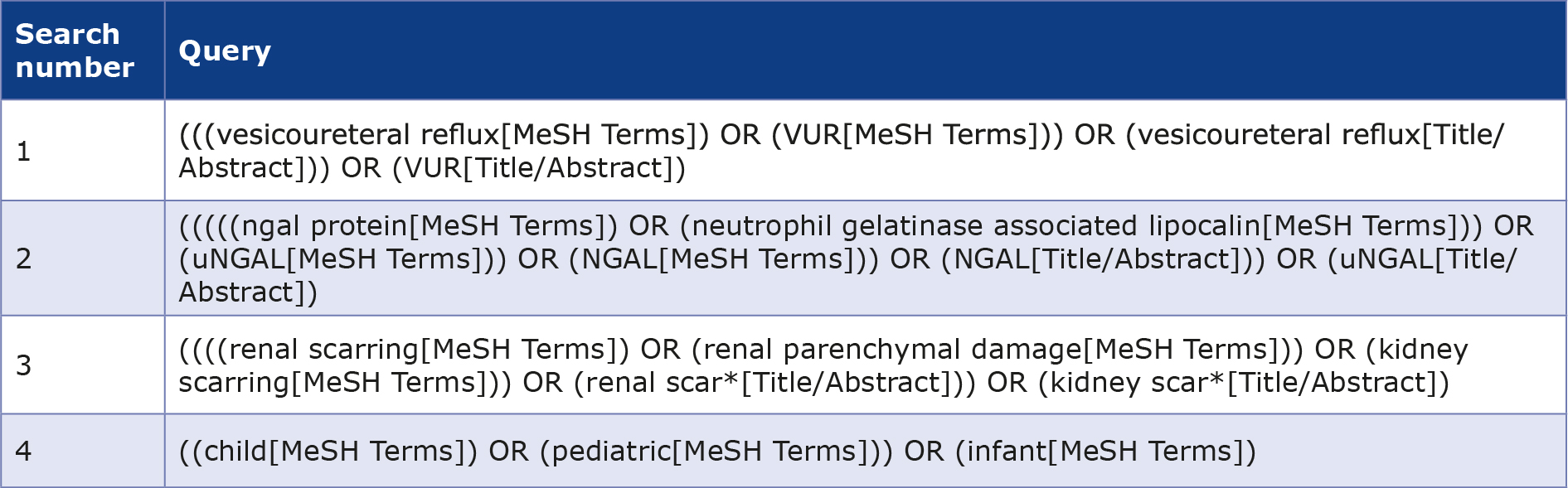
Whereas the Taylor & Francis online database we searched using the following strategy:
#1 vesicoureteral reflux or VUR,
#2 NGAL or neutrophil gelatinase associated lipocalin,
#3 renal scar or kidney scar or renal parenchymal damage,
#4 child or pediatric or infant.
Risk of bias assessment
The methodological quality of included studies was assessed using the QUADAS-2 (Quality Assessment of Diagnostic Accuracy Studies) tool [26]. This instrument evaluates risk of bias across 4 domains: patient selection, index test, reference standard, and flow and timing. Applicability concerns were also assessed for three domains.
Table 1. Literature search strategies in PubMed
Statistical analysis
Diagnostic accuracy data were synthesized using the MetaBayesDTA software (v1.5.2) [27]. From these models,pooled sensitivity, specificity, diagnostic odds ratio (DOR), positive likelihood ratio (LR+), and negative likelihood ratio (LR–) were derived, each with 95% confidence intervals (CI). The summary receiver operating characteristic (SROC) curve was generated to display overall test performance. Forest plots were constructed to illustrate sensitivity and specificity across individual studies.
Results
Data selection
The study selection process is summarized in the PRISMA flow diagram (Figure 1). Total of 127 studies were initially retrieved through the systematic search from 6 databases (PubMed, ScienceDirect, EuropePMC, OpenAlex, Taylor & Francis Online and Google Schoolar). After removing duplicates and ineligible studies by automation, 37 titles and abstracts were screened. From these, 7 full-text articles were evaluated for eligibility and then 3 studies excluded due to no data of sensitivity and specificity table (2 studies) and same data (1 study). There were 4 primary studies included in the final meta-analysis.
Figure 1. PRISMA flow diagram
Study characteristics
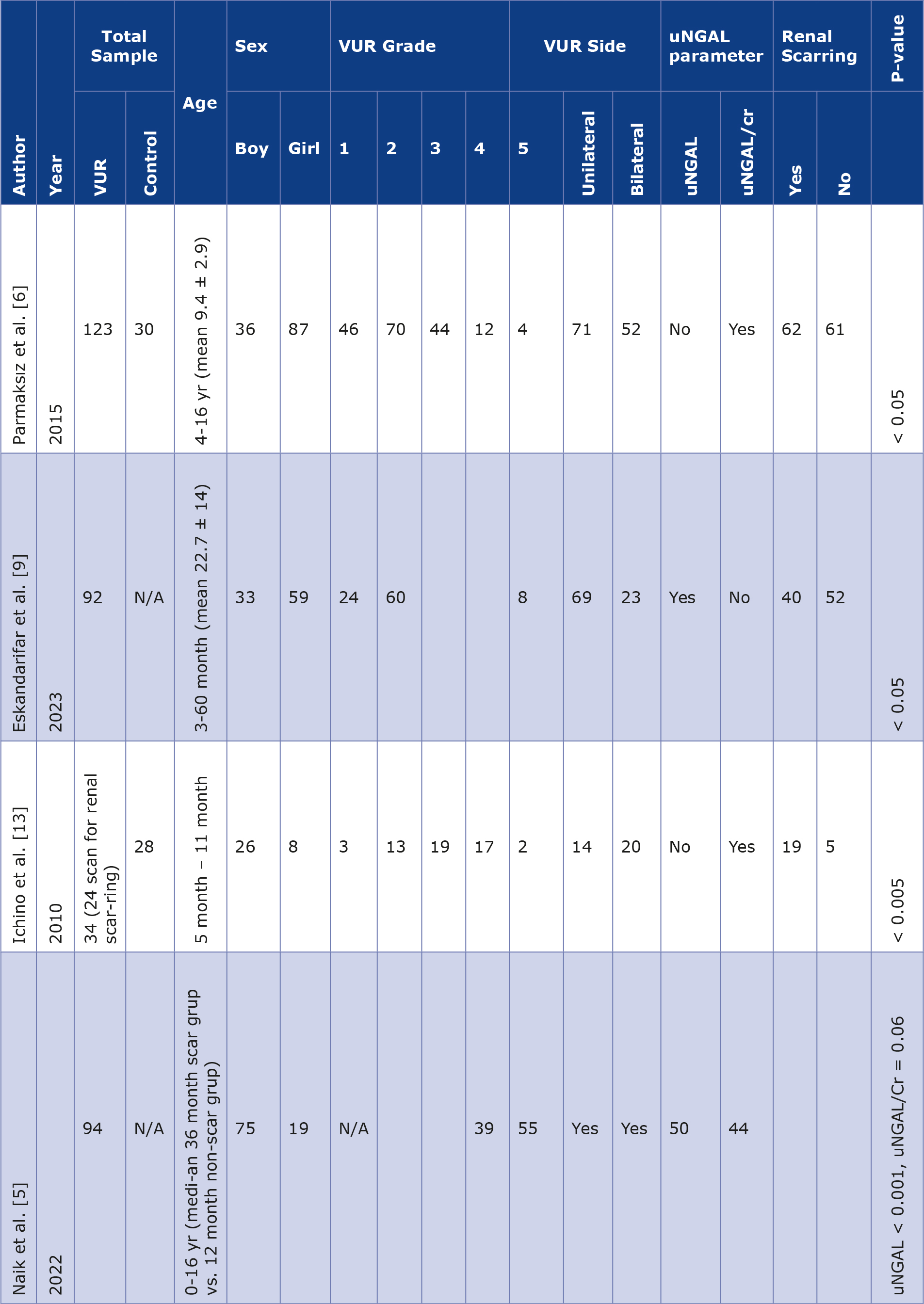
The four included studies were published between 2010 and 2023, enrolling between 34 and 123 pediatric patients with vesicoureteral reflux. Patient ages varied widely, from infants as young as 3 months to adolescents up to 16 years old. All studies included patients with varying grades of VUR (I to V). All studies used DMSA scintigraphy as the reference standard to confirm renal scarring, but there was variation in how urinary NGAL was measured and expressed. In some studies, NGAL was assessed as an absolute urinary concentration (uNGAL), while in others the values were normalized to urinary creatinine (uNGAL/Cr) to account for differences in hydration and urine output. 2 studies measured uNGAL and reported significant findings of analysis [5, 9]. 3 studies measured uNGAL/Cr with 2 studies had significant findings of analysis [6, 13] and 1 study was not significant findings of analysis (Table 2).
Table 2. Study characteristics
Diagnostic accuracy of uNGAL and uNGAL/Cr from each studies
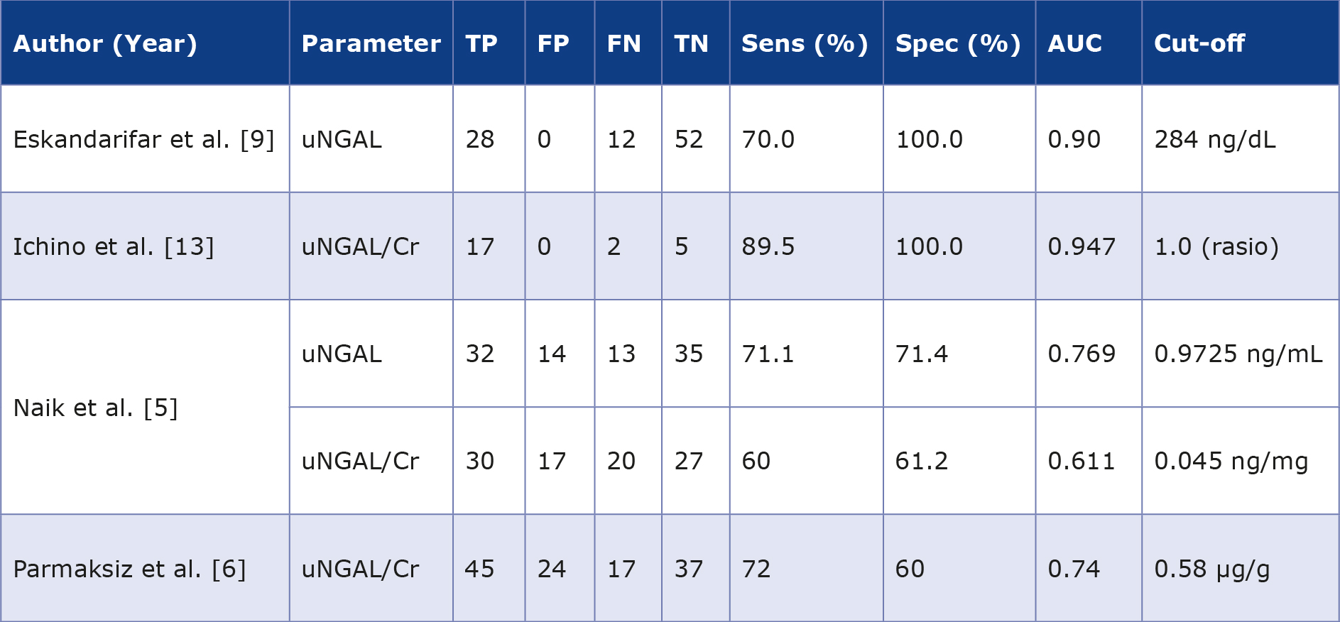
Eskandarifar et al. [9] conducted a cross-sectional study in 92 children with VUR. They measured absolute uNGAL levels and established a diagnostic threshold of 284 ng/dL. At this cut-off, the biomarker achieved a sensitivity of 70% and a specificity of 100% (Table 3), highlighting its potential as a highly specific indicator of renal scarring. The authors concluded that absolute uNGAL may be valuable for confirming scarring, but its moderate sensitivity limits its ability to exclude the condition.
Ichino et al. evaluated 34 pediatric patients and analyzed the uNGAL/Cr ratio. They reported excellent diagnostic performance, with sensitivity of 89.5% and specificity of 100% at a cut-off value of 1.0 (Table 3) [13]. This was one of the earliest studies to suggest that uNGAL could serve as a reliable noninvasive biomarker of renal scarring. However, there were 24 patients from 34 VUR patients that had renal scarring.
Table 3. Sensitivity and specificity data
Naik et al. evaluated 94 children with VUR, measuring both absolute uNGAL and the uNGAL/Cr ratio. Their findings showed that absolute uNGAL had moderate diagnostic ability (AUC 0.769, sensitivity 71.1%, specificity 71.4%) [5]. In contrast, the uNGAL/Cr ratio performed less well (AUC 0.611) (Table 3), suggesting that normalizing to creatinine did not improve diagnostic performance in this cohort.
Parmaksiz et al. carried out the largest study with 123 children. They focused on uNGAL/Cr and found moderate diagnostic accuracy, with sensitivity of 72% and specificity of 60% (Table 3) [6].
Pooled diagnostic accuracy of uNGAL
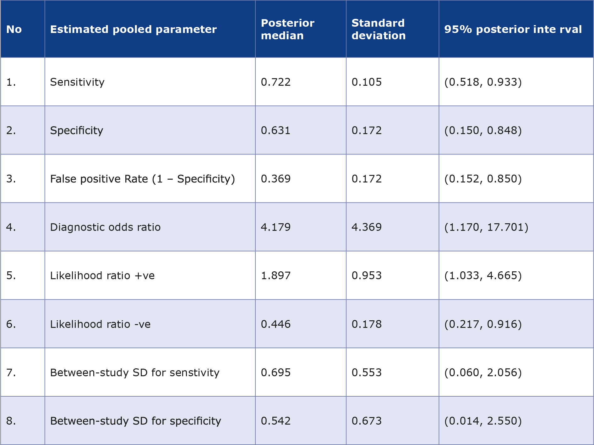
The analysis of uNGAL/Cr was conducted with MetaBayesDTA software as the only available parameter in thisstudy [27]. The analysis of uNGAL value parameters was not possible because 2 studies provided limited information about its diagnostic accuracy. When data from all 4 studies were synthesized, uNGAL/Cr demonstrated moderate diagnostic accuracy for detecting renal scarring (Table 4). The pooled sensitivity was 0.722 (SD 0.105; 95% CI: 0.518-0.933), while pooled specificity was 0.631 (SD 0.172; 95% CI: 0.150- 0.848). The overall diagnostic odds ratio was 4.179 (95% CI: 1.170-17.701), indicating more than fourfold higher odds of correctly identifying renal scarring when using uNGAL/Cr. The pooled positive likelihood ratio was 1.897 (95% CI: 1.033- 4.665), and the negative likelihood ratio was 0.446 (95% CI: 0.217-0.916), reflecting moderate rule-in but limited rule-out value. The false positive rate was 0.369 (95% CI: 0.152-0.850).
Table 4. Estimated parameter table
Quality assessment
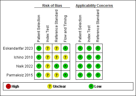
The quality assessment of the 4 included studies is presented in Figure 2. The QUADAS-2 (Quality Assessment of Diagnostic Accuracy Studies 2) tool was used and it revealed that all of 4 included studies had a moderate risk of bias.
Figure 2. Risk of bias analysis
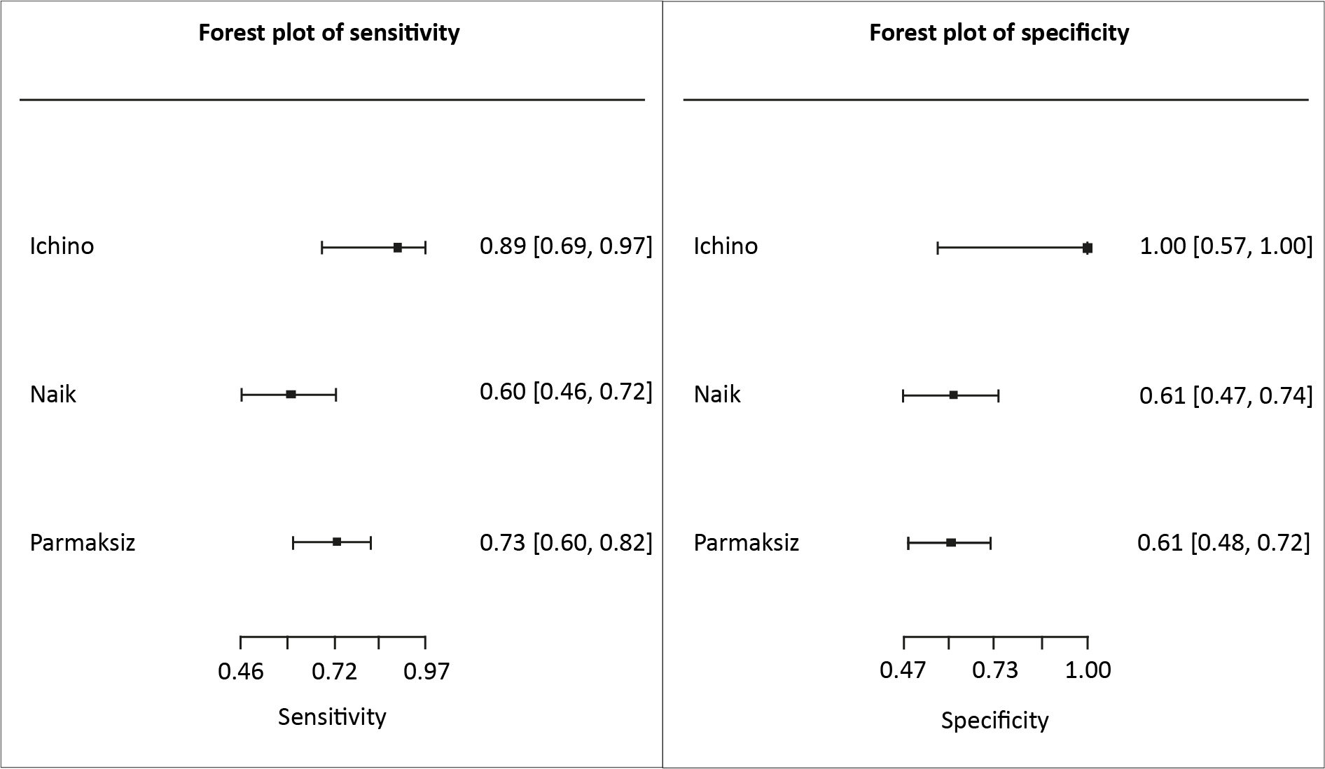
Forest plots of individual study estimated for sensitivity and specificity illustrate the variability across studies, with some studies reporting very high diagnostic accuracy while others demonstrated only moderate performance (Figure 3).
Figure 3. Sensitivity and specificity forest plot for uNGAL/Cr
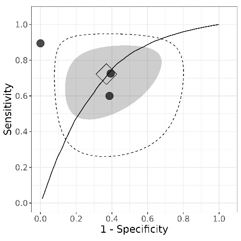
The summary receiver operating characteristic (SROC) curve indicated moderate overall discriminatory performance, with AUC values across studies ranging from 0.74 to 0.95. The credible intervals were relatively wide. The prediction region surrounding the HSROC was extensive, reflecting substantial heterogeneity across studies. The individual studies points were widely spread from summary curve that reinforce the variability diagnostic performance across the studies. Ichino et al. and Eskandarifar et al. [9, 13] plotted closer to the upper left corner of the curve, indicating high diagnostic power, while Naik et al. and Parmaksiz et al. [5-6] plotted closer to the midline, reflecting lower performance (Figure 4).
Figure 4. Summary receiver operating characteristic (SROC) plot
Discussion
The correlation between NGAL and renal scarring in VUR is multifaceted. NGAL is induced and secreted by renal tubular epithelial cells in response to various stressors, e.g. inflammation, ischemia, and injury, particularly within the context of UTIs and VUR [1, 6, 9, 15, 18].
The first mechanism contributing to this correlation is inflammatory response and bacteriostasis. NGAL functions as an acute-phase protein, showing significant increases during inflammation as in UTIs [6, 9, 14]. It plays a crucial role in innate immunity by sequestering iron from bacteria, thereby inhibiting their growth [10, 13-14]. In experimental pyelonephritis, NGAL gene and protein levels are upregulated in damaged renal tubule cells as part of this inflammatory response [6, 13].
The second mechanism is tubular cell injury and regeneration. NGAL is produced by injured renal tubular epithelial cells [14, 18-19, 23]. Its elevation in urine during renal scar formation is attributed to the regeneration process of renal tubular cells following injury [1, 13]. Initially, NGAL upregulation after renal insult may serve a protective function to limit injury [14]. However, persistent elevation of NGAL, particularly beyond the acute inflammatory phase (e.g. after 2 weeks post-pyelonephritis), indicates ongoing tubular injury (rather than just an active infection) and suggests sustained renal damage [9, 13, 15-16]. This sustained elevation can also predict the progression of CKD [14, 19].
The third mechanism is hypoxia and fibrosis. NGAL levels can be influenced by acute anemia and renal hypoxia, which may occur during acute pyelonephritis. NGAL has demonstrated a capacity to reduce renal hypoxia/reoxygenation-induced tubular cell damage, indicating a protective role [14]. Chronic inflammation, oxidative stress, and impaired tubular repair mechanisms (potentially exacerbated by renal scarring) contribute to a vicious cycle of injury and maladaptive repair, leading to tubulointerstitial damage and fibrosis associated with CKD [19, 23]. The concentration of NGAL is believed to reflect the ongoing kidney damage processes during CKD [15].
In the results of this study, there are 2 reported parameters: absolute urinary NGAL concentration (uNGAL) and urinary NGAL normalized to creatinine (uNGAL/Cr). Absolute uNGAL demonstrates higher specificity and better discrimination for renal scarring, as reported by Eskandarifar et al. and Naik et al. [5, 9]. However, absolute measurements are influenced by variations in urine dilution and hydration status [5].
Meta-analysis could only be performed for uNGAL/Cr because at least 3 studies reported comparable data. Absolute uNGAL values could not be performed because of small number of studies that reported comparable data. Overall, absolute uNGAL appears superior for ruling in scarring, whereas uNGAL/Cr offers theoretical adjustment for the dilution effects but lower diagnostic accuracy.
Our study demonstrated that uNGAL/Cr has a moderate diagnostic value in detecting renal scarring in pediatric VUR when compared with DMSA scintigraphy. Our pooled results showed sensitivity of 72.2% and specificity of 63.1%, with a diagnostic odds ratio of 4.18, indicating that uNGAL/Cr may serve as a supportive biomarker but not a standalone replacement for imaging.
The reason for such findings is the highly varied creatinine excretion in children (influenced by age, muscle mass, and growth), which undermines the ratio’s reliability [1]. Parmaksiz et al. found moderate accuracy for uNGAL/Cr (AUC 0.74; sensitivity 72%, specificity 60%), while Naik et al. reported poor performance (AUC 0.611; sensitivity 60%, specificity 61.2%) [5-6]. In contrast, Ichino et al. reported strong diagnostic value of uNGAL/Cr (AUC 0.947; sensitivity 89.5%, specificity 100%), highlighting the inconsistency across studies [13].
Although NGAL does not match the diagnostic precision of DMSA, its ability to reflect renal tubular injury at the molecular level provides a theoretical advantage in identifying early damage before structural changes appear on scintigraphy [11]. Thus, uNGAL may be valuable as a predictor biomarker, reducing the number of DMSA scans and cumulative radiation exposure. Ichino et al. and Eskandarifar et al. highlighted the high specificity of uNGAL (100%), suggesting its role as a confirmatory tool in identifying patients with renal scarring [9, 13]. However, the limited sensitivity underscores that uNGAL is insufficient to exclude renal scarring.
A critical consideration in interpreting uNGAL accuracy is its biological nature as a real-time marker of active tubular stress. While DMSA scintigraphy cannot structurally differentiate between a static, congenital dysplastic lesion (arising from prenatal VUR) and an acquired post-inflammatory scar (arising from febrile UTIs), uNGAL may offer functional differentiation. Congenital renal dysplasia without ongoing infection represents a fixed structural defect where ongoing tubular injury is minimal, theoretically yielding lower uNGAL/ Cr ratios. In contrast, post-pyelonephritic scar formation involves an active, protracted tubulointerstitial inflammatory response that significantly upregulates NGAL expression [23]. Thus, uNGAL’s clinical utility may lie not just in replacing imaging, but as a biomarker for distinguishing active, progressive post-infectious reflux nephropathy versus stable, congenital dysplastic parenchyma.
Other urinary biomarkers (e.g. kidney injury molecule-1 (KIM-1)), have also been explored. Some authors reported associations between KIM-1 and renal scarring, while others did not find significant differences [11-12]. Given these inconsistencies, combining NGAL with other markers may enhance diagnostic accuracy. Ganapathy et al. demonstrated that machine learning models integrating NGAL, KIM-1, and creatinine had superior performance (AUC 0.83) compared to individual biomarkers [4]. This suggests that multi-marker strategies may represent the future of non-invasive diagnostics in pediatric nephrology.
Our analysis is limited by the fact that only 3 studies were included, therefore the pooled estimates were inconsistent. The difference between definition and calculation of cut-offs across studies probably have contributed to the heterogeneity. In addition, the wide range of between-study SD for sensitivity and specificity adds heterogeneity to the result of polledestimates. However, because of the limited number of studies, exploration of heterogeneity through subgroup analysis or meta-regression was not feasible. Therefore, our findings should be interpreted with caution and their generalizability to other settings remains limited.
Future research should focus on standardizing cut-off definition and calculation for both uNGAL and uNGAL/Cr, validating serum NGAL alongside urinary NGAL, and evaluating the role of multi-marker panels in large, multicenter cohorts. Longitudinal studies should also explore NGAL’s utility in monitoring disease progression and treatment response, offering potential for more personalized and less invasive management of pediatric VUR.
Conclusion
Urinary NGAL, particularly the uNGAL/Cr demonstrates moderate diagnostic value for detecting renal scarring in pediatric VUR when compared with scintigraphy. While uNGAL may not replace DMSA in early diagnosis, it offers potential as a non-invasive additional biomarker to reduce radiation exposure. Future research about uNGAL’s diagnostic value should focus on large-scale, multi-center prospective studies with standardized cut-off values. Additionally, longitudinal studies may be helpful in detecting early renal deterioration.
Funding
Self funded.
Conflict of interest
None.
References
| 1. |
Colceriu M-C, Aldea PL, Boț (Răchişan) A-L, Bulată B, Delean D, Grama A, et al. The Utility of Noninvasive Urinary Biomarkers for the Evaluation of Vesicoureteral Reflux in Children. Int J Mol Sci [Internet]. 2023;24(24):17579. Available from: https://www.mdpi.com/1422-0067/24/24/17579.
|
| 2. |
Mattoo TK. Vesicoureteral Reflux and Reflux Nephropathy. Adv Chronic Kidney Dis [Internet]. 2011;18(5):348–54. Available from: https://linkinghub.elsevier.com/retrieve/pii/S1548559511001200.
|
| 3. |
Putri UMA, Raharja PAR, Situmorang GR, Wahyudi I, Rodjani A, Puspitasari HA, et al. Biomarker for renal scarring screening in children with vesicoureteral reflux: a systematic review. Front Pediatr [Internet]. 2025;13. Available from: https://www.frontiersin.org/articles/10.3389/fped.2025.1621716/full.
|
| 4. |
Ganapathy S, K.T. H, Jindal B, Naik PS, Nair N. S. Comparison of diagnostic accuracy of models combining the renal biomarkers in predicting renal scarring in pediatric population with vesicoureteral reflux (VUR). Irish J Med Sci (1971 -) [Internet]. 2023;192(5):2521–6. Available from: https://link.springer.com/10.1007/s11845-023-03275-z.
|
| 5. |
Naik PB, Jindal B, Kumaravel S, Halanaik D, Rajappa M, Naredi BK, et al. Utility of Urinary Biomarkers Neutrophil Gelatinase-Associated Lipocalin and Kidney Injury Molecule-1 as a Marker for Diagnosing the Presence of Renal Scar in Children with Vesicoureteral Reflux (VUR). J Indian Assoc Pediatr Surg [Internet]. 2022;27(1):83–90. Available from: https://journals.lww.com/10.4103/jiaps.JIAPS_334_20.
|
| 6. |
Parmaksız G, Noyan A, Dursun H, İnce E, Anarat R, Cengiz N. Role of new biomarkers for predicting renal scarring in vesicoureteral reflux: NGAL, KIM-1, and L-FABP. Pediatr Nephrol [Internet]. 2016;31(1):97–103. Available from: http://link.springer.com/10.1007/s00467-015-3194-3.
|
| 7. |
Aeddula N, Baradhi K. Reflux Nephropathy. In: StatPearls [Internet] [Internet]. Treasure Island (FL): StatPearls Publishing; 2026. Available from: https://www.ncbi.nlm.nih.gov/books/NBK526055/.
|
| 8. |
Prasad MM, Cheng EY. Imaging studies and biomarkers to detect clinically meaningful vesicoureteral reflux. Investig Clin Urol [Internet]. 2017;58(Suppl 1):S23. Available from: https://icurology.org/DOIx.php?id=10.4111/icu.2017.58.S1.S23.
|
| 9. |
Eskandarifar A, Naghshizadian R, Tari A. Assessment of Urinary Level of Neutrophil Gelatinaseassociated Lipocalin (NAGL) in Children with Renal Scar Due to Vesicoureteral Reflux. Iran J Kidney Dis [Internet]. 2023;17(1):14–9. Available from: http://www.ncbi.nlm.nih.gov/pubmed/36739486.
|
| 10. |
ElNahal ASM, Ibrahim A, Abdelhalim E, Essawy SH, Behiry A, Saadoun M, et al. Serum neutrophil gelatinase associated Lipocalin as a novel biomarker for diagnosing acute pyelonephritis in adults. Sci Rep [Internet]. 2025;15(1):19651. Available from: https://www.nature.com/articles/s41598-025-03646-9.
|
| 11. |
Kostic D, Beozzo GPNS, do Couto SB, Kato AHT, Lima L, Palmeira P, et al. The role of renal biomarkers to predict the need of surgery in congenital urinary tract obstruction in infants. J Pediatr Urol [Internet]. 2019;15(3):242.e1-242.e9. Available from: https://linkinghub.elsevier.com/retrieve/pii/S1477513119300671.
|
| 12. |
Yiğit D, Taşkınlar H, Avlan D. Can serum Neutrophil Gelatinase Associated Lipocalin and Kidney Injury Molecule−1 help in decision making for surgery in antenatally dedected hydronephrosis. J Pediatr Urol [Internet]. 2021;17(1):71.e1-71.e7. Available from: https://linkinghub.elsevier.com/retrieve/pii/S1477513120305702.
|
| 13. |
Ichino M, Kusaka M, Kuroyanagi Y, Mori T, Morooka M, Sasaki H, et al. Urinary Neutrophil-Gelatinase Associated Lipocalin is a Potential Noninvasive Marker for Renal Scarring in Patients With Vesicoureteral Reflux. J Urol [Internet]. 2010;183(5):2001–7. Available from: http://www.jurology.com/doi/10.1016/j.juro.2010.01.031.
|
| 14. |
Lee JH, Yim HE, Yoo KH. Associations of Plasma Neutrophil Gelatinase-associated Lipocalin, Anemia, and Renal Scarring in Children with Febrile Urinary Tract Infections. J Korean Med Sci [Internet]. 2020;35(10). Available from: https://jkms.org/DOIx.php?id=10.3346/jkms.2020.35.e65.
|
| 15. |
Gavrilovici C, Dusa CP, Iliescu Halitchi C, Lupu VV, Spoiala EL, Bogos RA, et al. The Role of Urinary NGAL in the Management of Primary Vesicoureteral Reflux in Children. Int J Mol Sci [Internet]. 2023;24(9):7904. Available from: https://www.mdpi.com/1422-0067/24/9/7904.
|
| 16. |
Forster CS, Devarajan P. Neutrophil gelatinase-associated lipocalin: utility in urologic conditions. Pediatr Nephrol [Internet]. 2017;32(3):377–81. Available from: http://link.springer.com/10.1007/s00467-016-3540-0.
|
| 17. |
Moon JH, Yoo KH, Yim HE. Urinary neutrophil gelatinase-associated lipocalin: a marker of urinary tract infection among febrile children. Clin Exp Pediatr [Internet]. 2021;64(7):347–54. Available from: http://e-cep.org/journal/view.php?doi=10.3345/cep.2020.01130.
|
| 18. |
Bagińska J, Korzeniecka-Kozerska A. Are Tubular Injury Markers NGAL and KIM-1 Useful in Pediatric Neurogenic Bladder? J Clin Med [Internet]. 2021;10(11):2353. Available from: https://www.mdpi.com/2077-0383/10/11/2353.
|
| 19. |
Srivast A, Anand S, Kumar H, Meena JK, Verma A, Luthra K, et al. Urinary NGAL and Renalase as Non-Invasive Biomarkers for Detection of Deterioration of Kidney Function and Kidney Scarring in Children with Neurogenic Bladder [Internet]. 2025. Available from: https://www.researchsquare.com/article/rs-5998098/v1.
|
| 20. |
Zhang Y, Chen C, Mitsnefes M, Huang B, Devarajan P. Evaluation of diagnostic accuracy of urine neutrophil gelatinase-associated lipocalin in patients with symptoms of urinary tract infections: a meta-analysis. Front Pediatr [Internet]. 2024;12. Available from: https://www.frontiersin.org/articles/10.3389/fped.2024.1368583/full.
|
| 21. |
Jagadesan I, Agarwal I, Chaturvedi S, Jose A, Sahni R, Fleming J. Urinary neutrophil gelatinase associated lipocalin – A sensitive marker for urinary tract infection in children. Indian J Nephrol [Internet]. 2019;29(5):340. Available from: https://indianjnephrol.org/urinary-neutrophil-gelatinase-associated-lipocalin-a-sensitive-marker-for-urinary-tract-infection-in-children/.
|
| 22. |
Nickavar A, Valavi E, Safaeian B, Moosavian M. Validity of urine neutrophile gelatinase-associated lipocalin in children with primary vesicoureteral reflux. Int Urol Nephrol [Internet]. 2020;52(4):599–602. Available from: http://link.springer.com/10.1007/s11255-019-02355-3.
|
| 23. |
Gkiourtzis N, Stoimeni A, Michou P, Charitakis N, Cheirakis K, Liakos V, et al. The value of procalcitonin and urinary NGAL in the prediction of acute pyelonephritis and kidney scarring in pediatric patients with a history of febrile urinary tract infection: a systematic review and meta-analysis. Pediatr Nephrol [Internet]. 2026;41(2):323–37. Available from: https://link.springer.com/10.1007/s00467-025-06885-0.
|
| 24. |
Sadr Moharerpour S, Otukesh H, Hoseini Shamsabadi R, Ghorbani H, Nakhaiee S, Seirafianpour F, et al. Assessment of Urinary and Serum Neutrophil Gelatinase-Associated Lipocalin (NGAL) Levels as Novel Predictors for Vesicoureteral Reflux Diagnosis in Children with Febrile Urinary Tract Infection. Med J Islam Repub Iran [Internet]. 2024; Available from: https://mjiri.iums.ac.ir/article-1-8206-en.html.
|
| 25. |
Adams DP, Gozlan EC, Medikonda N, Song JJ, Sahoo A, Yeagley M, et al. Stratification of Wilms tumor patients using physicochemical properties of the adaptive immune receptor polypeptides, IGL and TRG. J Pediatr Urol [Internet]. 2025;21(1):130–5. Available from: https://linkinghub.elsevier.com/retrieve/pii/S1477513124005333.
|
| 26. |
Whiting PF, Rutjes AWS, Westwood ME, Mallett S, Deeks JJ, Reitsma JB, et al. QUADAS-2: A Revised Tool for the Quality Assessment of Diagnostic Accuracy Studies. Ann Intern Med [Internet]. 2011;155(8):529–36. Available from: https://www.acpjournals.org/doi/10.7326/0003-4819-155-8-201110180-00009.
|
| 27. |
Cerullo E, Sutton AJ, Jones HE, Wu O, Quinn TJ, Cooper NJ. MetaBayesDTA: codeless Bayesian meta-analysis of test accuracy, with or without a gold standard. BMC Med Res Methodol [Internet]. 2023;23(1):127. Available from: https://bmcmedresmethodol.biomedcentral.com/articles/10.1186/s12874-023-01910-y.
|